Download
1 / 20
211 likes | 649 Vues
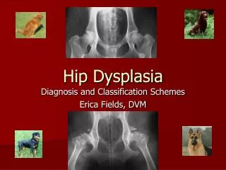
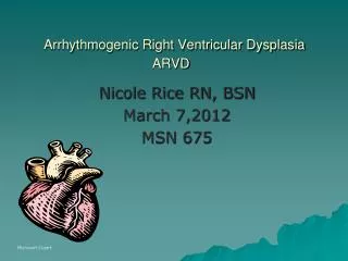
Arrhythmogenic Right Ventricular Dysplasia ARVD. Nicole Rice RN, BSN March 7,2012 MSN 675. Microsoft Cipart. Case Study. Back to question.

E N D
Arrhythmogenic Right Ventricular Dysplasia ARVD Nicole Rice RN, BSN March 7,2012 MSN 675 MicrosoftCipart
Case Study Back to question Lorenzo Guiterrez, 39, was driving when he experienced sudden onset of chest pains, dizziness, diaphoresis. He happened to be driving by a local emergency department. As he entered, he collapsed. He was revived after CPR and defibrillation, restoring his ventricular tachycardia (VT) to normal sinus rhythm (NSR). He reported having feelings like this before, and two of his family members had died of sudden cardiac death. His past medical history was significant for mild hypertension. “ARVD/C is a leading cause of sudden death among young athletes, although people within a broad range of ages and activity levels have this condition. It affects men and women of all races, and approximately 1 out of every 5,000 is diagnosed with ARVD/C. It’s possible to have this disease without knowing it. These silent carriers may not recognize its symptoms when—or if –they occur. Palpitations, fainting, chest pain, and rapid heartbeat are some warning signals of ARVD/C.” (2010 Johns Hopkins Patient Education Brochure, p. 1)
Tutorial Objectives Define Arrhythmogenic Right Ventricular Dysplasia (ARVD) Identify common signs and symptoms in the physical exam of the ARVD patient Outline commonly used diagnostic tests to confirm a ARVD diagnosis Describe treatments used to help those who suffer from ARVD
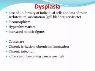
What is ARVD? ARVD is a dangerous cardiac condition that can cause irregular heart rhythms and sudden death. It is characterized by a thinned and dilated right ventricle (RV) from a progressive loss of myocyteswhich are replaced with fibrous and fatty tissue in the RV myocardium. The fatty tissue disrupts electrical pathways. Fatty tissue in RV causes abnormal contractions like ventricular tachycardia (VT) thus abnormal ejection of blood.
The Johns Hopkins Arrhythmogenic Right Ventricular Dysplasia Program has described that it is widely recognized that ARVD is often caused by mutations in proteins called desmosomal proteins. These proteins are like a bridge that links one heart cell to the next. Major components of desmosomes are plakophilin-2, desmoglein -2, desmocollin-2, desmoplakin, and plakoglobin. It has been discovered that those who are afflicted with ARVD have genetic abnormalities in the genes encoding for these desmosomal proteins. Since the mechanical bonds are weak and faulty, over time and exertion, the process of pulling apart causes formation of scarring and fat replacement. We would see the pulling action when the heart is being worked, like during exercise. Microsoft Clipart
What is ARVD? • The condition is precipitated by a release of catecholamine surges like exercise, dehydration, stress, cocaine or electrolyte imbalances. • Clinical manifestations include palpitations, syncope, cardiac arrest, mental confusion, anxiety. In progressive phases with left ventricular involvement dyspnea on exertion and orthopnea are present. Microsoft Clipart
Four ARVD Stages • Concealed phase: Pt usually asymptomatic, may have minor VT, RV is only seen with subtle changes. These clients are at high risk of sudden death. • Overt phase: Noted structural and functional changes in the RV. Symptoms seen are ventricular dysrhythmias, presyncope, syncope, and palpitiations. • Weakening of RV: The RV has dilated and weakened. Client exhibits symptoms like RV failure including edema of lower extremities, abdominal distention, dyspepsia, and anorexia. • Weakening of the LV: The LV dilates and weakens. Now see symptoms of heart failure which include dyspnea on exertion, orthopnea, and breathlessness. Source: Andrews, T., Cook, S., Baumeister, M., Hickey, K., (2010). Sudden Killer: Arrhythmogenic right ventricular cardiomyopathy. Nursing, 40 (7) p. 7-11.
Question about topic Note: there is more than one right answer Palpitations, chest pain, irregular heart rhythm Yes that’s it! Mental confusion, anxiety You bet! Lower extremity edema, large abdominal girth Sorry, those are later signs After reviewing the case study, what are the early signs and symptoms of ARVD? Click here to see case study.
Testing your knowledge so far Note: there is more than one right answer Happens to young athletes Yes, you are correct Clients can present at different stages of ARVD Yes, four stages of ARVD Fatty fibrous tissue in RV Yes, that is right Knowing what you have already seen, what are the characteristics of ARVD?
ECG abnormalities: • 90% of patients have some abnormality, typically T wave inversions. Also noted is a right bundle branch block. • “In addition, an epsilon wave secondary to slowing of intraventricular conduction. This wave is seen at the terminal notch in the QRS complex, and most often seen on signal-averaged ECG, (Andrews,Cook,Baumeister,Hickey (2010), p. 4.)”. The epsilon wave is also described as “distinct waves of small amplitude present in the ST segment in right precordial leads, (Yerra,Caskey,Modi,Reddy (2008), p. 4)”. • Also noted are a wide QRS greater than 110 milliseconds and a prolonged S wave in leads V1 to V3 due to delayed RV repolarization.
Powered by Blogger. 2 of 3 Illustration of epsilon wave Red triangles mark epsilon wave, also note inverted T wave. wikipedia image used under creative commons license
Cross section of heart showing pronounced adipose infiltration of right ventricular free wall and nearly normal left ventricle and ventricular septum. Image used with permission from Johns Hopkins ARVD Dysplasia Program. http://www.ncbi.nlm.nih.gov/sites/ppmc/articles/PMC483655 Fatty infiltrate
Diagnostic Testing • ECG, Holter monitoring, Echocardiograph, Cardiac MRI, and RV angiography, cardiac biopsy. • Additional test that is helpful is ECG with exercise stress testing. This helps to see with stress on the heart the frequency and duration of ectopic beats or ventricular dysrhythmias. Microsoft Clipart
Diagnostic Findings • Echocardiographic abnormalities: RV dilation, hypokinesis, and very thin RV free wall. Tricuspid regurgitation may also be noted. • MRI: RV-free wall changes. Fatty infiltration, thinning, and akinesis of RV free-wall are commonly noted. • Angiography: “akinetic or dyskenetic bulging may be localized in the triad of dysplasia areas (infundibular, apical, and subtricuspid) this test has 90% specificity (Andrews,Cook,Baumeister,Hickey (2010), p. 4.) “. “For a clinician to arrive at a definitive diagnosis of ARVD, two major criteria or one major and two minor critieria must be present, or four minor criteria from different groups (Andrews,Cook,Baumeister,Hickey (2010), p. 4.)”.
Review Note: there is more than one right answer the epsilon wave definitely, that is a hallmark sign T wave inversion yes! Prolonged S wave Yes! Also wide QRS Thinking back to the case study, what kinds of ECG findings might we see in diagnostic studies after he has stabilized?
Genetic Testing • ARVD is an inherited disease with variable presentation within families. Studies about the genetic possibilities of the disease found that nearly 40% of people with familial tendencies have ARVD. It is primarily an autosomal dominant genetic disorder, or a 50% chance that offspring will develop ARVD or its symptoms. • A genetic mutation in the protein plakophilin-2 (PKP2) is associated with up to 45% of ARVD patients. • Patients with PKP2 are also more likely to develop ARVD at an early age. Further testing has been taking place to isolate more genes that may identify a person at risk for developing ARVD. (Yerra,Caskey,Modi,Reddy (2008), p.6), (Andrews,Cook,Baumeister,Hickey (2010), p.4)http://www.hopkinsmedicine.org/hmn/F07/medical.cfm Microsoft Clipart
Treatment of ARVD Medications: • Sotalol (beta-adrenergic blocker) • Amiodarone (anti-arrhythmic) • Warfarin – “prevents embolic development secondary to high residual blood volume in RV (Andrews,Cook,Baumeister,Hickey (2010), p. 4.)”
Treatment of ARVD • ICD placement – commonly performed, with caution due to thinning of RV wall causing the risk of perforation. • Radio Frequency Ablation - for those with less risk of perforation it is a therapeutic option in patients unresponsive or intolerant to antiarrhythmic drugs. In recent research, (Arbelo, 2010) catheter ablation should be the first line approach in patient with recurrent monomorphic ventricular tachycardia. This will disable the malfunctioning circuits and reduce incidence of arrhythmia occurring.
Now that I know about ARVD… It is very important that a thorough physical assessment is performed, especially for young adults undergoing physicals for competitive or contact sports. The assessment should include asking questions of parents about: ● family history of palpitations ● lightheadedness ● blackouts or syncopal events ● evaluation of ECG if history or symptoms are present ● If there are concerns or a strong history of sudden cardiac events (arrhythmias, arrest, MI) in close relatives a family pedigree should be obtained with at least three generations.
Literature Cited Andrews, T., Cook, S., Baumeister, M., Hickey, K., (2010). Sudden Killer: Arrhythmogenic right ventricular cardiomyopathy. Nursing, 40 (7) p. 7-11. Arbelo, E., Josephson, M., (2010). Ablation of ventricular arrhythmias in arrhythmogenic right ventricular dysphasia. Journal of Cardiovascular Electrophysiology, 21 (4): 473-86. Porth, C. M., & Matfin, G., (2009). Pathophysiology: Concepts of altered health states (8th ed.). Philadelphia, PA: Lippincott Williams & Wilkins. http://en.wikipedia.org/wiki/ARVD retrieved March 29, 2012. Yerra, L., Caskey, D., Modi, K., Reddy, P., (2008). Arrhythmogenic Right Ventricular Dysplasia/Cardiomyopathy: Cinical Profile of Four Patients and Review. Southern Medical Journal, 101 (3) p. 309-316. Johns Hopkins Medicine, ARVD Program Patient Brochure, January 2010, Authored by Johns Hopkins Medical Professionals. Images utilized with permission from Johns Hopkins ARVD Program official. http://www.arvd.com