Introduction to Affymetrix GeneChip data
710 likes | 1.11k Vues
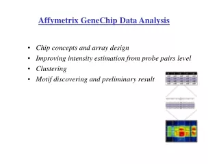
Introduction to Affymetrix GeneChip data. Stat 246, Spring 2002, Week 16. Summary. Review of technology Probeset summaries What we do: our 4 steps Assessing the technology and the different expression measures How robustness works. *. *. *. *. *. Probe arrays. Hybridized Probe Cell.

Introduction to Affymetrix GeneChip data
E N D
Presentation Transcript
Introduction to Affymetrix GeneChip data Stat 246, Spring 2002, Week 16
Summary • Review of technology • Probeset summaries • What we do: our 4 steps • Assessing the technology and the different expression measures • How robustness works
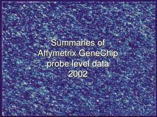
* * * * * Probe arrays Hybridized Probe Cell GeneChipProbe Array Single stranded, labeled RNA target Oligonucleotide probe 24m Millions of copies of a specific oligonucleotide probe 1.28cm >200,000 different complementary probes Image of Hybridized Probe Array Compliments of D. Gerhold
Image analysis • About 100 pixels per probe cell • These intensities are combined to form one number representing expression for the probe cell oligo • Possibly room for improvement
Summarize 20 PM,MM pairs (probe level data) into one number for each probe set (gene) We call this number an expression measure Affymetrix GeneChip Software has defaults. Does it work? Can it be improved? The big picture
Where is the evidence that it works? Lockhart et. al. Nature Biotechnology 14 (1996)
Comments • The chips used in Lockhart et. al. contained around 1000 probes per gene • Current chips contain 11-20 probes per gene • These are quite different situations • We haven’t seen a plot like the previous one for current chips
Some possible problems What if • a small number of the probe pairs hybridize much better than the rest? • removing the middle base does not make a difference for some probes? • some MMs are PMs for some other gene? • there is need for normalization? We explore these possibilities using a variety of data sets
Competing measures of expression • GeneChip® older software uses Avg.diff with A a set of suitable pairs chosen by software. 30%-40-% can be <0. • Log PMj/MMj was also used. • For differential expression Avg.diffs are compared between chips.
Competing measures of expression, 2 • Li and Wong fit a model They consider i to be expression in chip i • Efron et al consider log PM - 0.5 log MM. It is much less frequently <0. • Another summary is the second largest PM, PM(2)
Competing measures of expression, 3 • GeneChip® newest version uses something else, namely with MM* a version of MM that is never bigger than PM.
Competing measures of expression, 4 • Why not stick to what has worked for cDNA? Again A is a suitable set of pairs. Care needed with BG, and we need to robustify.
What we do: four steps We use only PM, and ignore MM. Also, we • Adjust for background on the raw intensity scale • Take log2 of background adjusted PM • Carry out quantile normalization of log2(PM-BG), with chips in suitable sets • Conduct a robust multi-chip analysis (RMA) of these quantities We call our approach RMA
Why remove background? White arrows mark the means
Background model: pictorially + = Signal + Noise = Observed
Background model: formulae • Observed PM intensity denoted by S. • Model S as the sum of a signal X and a background Y, S=X+Y, where we assume X is exponential () and Y is Normal (, 2), X, Y independent random variables. • Background adjusted values are then E(X|S=s), which is a + b[(a/b) - ((s-a)/b)]/[(a/b) - ((s-a)/b) - 1], where a = s - - 2 , b = , and and are the normal density and cumulative density, respectively. This is our model and formula for background correction.
Observed PM vs Corrected PM As s increases, the background correction asymptotes to s - - 2 . In practice, >> 2, so this is ~ s - .
Quantile normalization • Quantile normalization is a method to make the distribution of probe intensities the same for every chip. • The normalization distribution is chosen by averaging each quantile across chips. • The diagram that follows illustrates the transformation.
Quantile normalization: in words • The two distribution functions are effectively estimated by the sample quantiles. • Quantile normalization is fast • After normalization, variability of expression measures across chips reduced • Looking at post-normalization PM vs pre-normalization PM (natural and log scales), you can see transformation is non linear.
Quantile normalization: formulae Normalization distribution F2(x) Raw data xnorm = F2-1(F1(x)) Density function Distribution function F1(x)
Dilution series: before and after quantile normalization in groups of 5 Note systematic effects of scanners 1,…,5 in before boxplots
Normalization reduces variability in comparison with Quantile vs Un-normalized Quantile vs Affymet. normalized Vertical: log[var q. norm/var other]; Horizontal: Aver. log mean Note differences in vertical scales
Probe effects: spike-in experiments • Set A: 11 control cRNAs were spiked in, all at the same concentration, which varied across chips. • Set B: 11 control cRNAs were spiked in, all at different concentrations, which varied across chips. The concentrations were arranged in 12x12 cyclic Latin square (with 3 replicates)
RMA = Robust multi-chip analysis • Background correct PM • Normalize (quantile normalization) • Assume additive model: • Estimate chip effects ai and probe effects bjusing a robust method
Comparing expression summaries using spike-in data Later we consider 23 different combinations of concentrations
Dilution experiment • cRNA hybridized to human chip (HGU95) in range of proportions and dilutions • Dilution series begins at 1.25 g cRNA per GeneChip array, and rises through 2.5, 5.0, 7.5, 10.0, to 20.0 g per array. 5 replicate chips were used at each dilution • Normalize just within each set of 5 replicates • For each probe set compute expression, average and SD over replicates