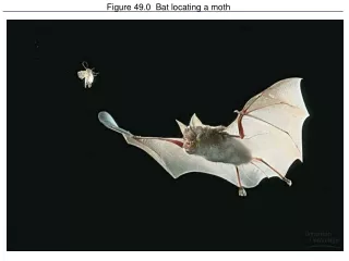
Figure 49.0 Bat locating a moth
820 likes | 1.05k Vues
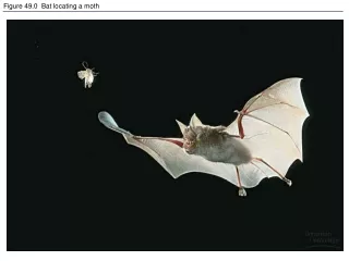
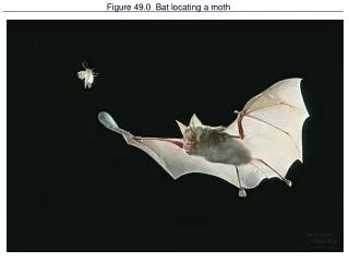
Figure 49.0 Bat locating a moth. Sensory receptors transduce stimulus energy and transmit signals to the nervous system. Four Basic Processes Must Occur For You To Sense The Environment Sensory Transduction Amplification Transmission Integration.

Figure 49.0 Bat locating a moth
E N D
Presentation Transcript
Sensory receptors transduce stimulus energy and transmit signals to the nervous system. • Four Basic Processes Must Occur For You To Sense The Environment • Sensory Transduction • Amplification • Transmission • Integration
Umm, Umm, Good. . . Taste Receptors • Taste buds are not as localized on the tongue like we once thought. • But a sweet, bitter, sour, salty, molecule binds to its particular receptor. • Signal is amplified and sets off a Signal Transduction Pathway • Potassium ion channels will be closed by the action of second messengers • Sodium moves into the cell causing depolarization. • This causes calcium ion uptake into the receptor (like at the end of the presynaptic neuron) • Neurotransmitters are released that stimulate a sensory neuron and subsequent action potential.
Figure 49.6 Specialized electromagnetic receptors: Rattle snake with infrared receptors, beluga whale pod Infrared receptors: sense infrared radiation given off by prey (mouse or jogger’s ankle)
Side with no screening pigment Figure 49.7 Eye cups and orientation behavior of a planarian Side with no screening pigment
Figure 49.8 Compound eyes (a) Image Forming Found in insects and crustaceans Consists of lots of ommatidia Each ommatidia has its own lens to focus light Differences in light intensity produce a mosaic image. Detects movement
Figure 49.8x2 Insect vision: A black-eyed Susan (Rudbeckia hirta) as humans see it and in ultraviolet light as visible to an insect
Forms the cornea Figure 49.9 Structure of the Vertebrate Eye Forms the iris Highest Conc. Of Cones No photoreceptors: “blind spot”
Figure 49.10 Focusing in the mammalian eye When ciliary muscles contract, the suspensory ligaments relax and allow the lens to “round.”
The Biochemistry of Sight • Rods are responsible for noncolor dim light; cones are responsible for color vision in bright light. • Rod photoreceptor: made up of outer and inner segments, cell body, synaptic region where the nerve impulse is passed to a retinal nerve cell. • Outer Segment: made up of stacks of discs containing rhodopsin molecules. The rhodopsin contains a molecule called retinal and opsin that can actually exist in two forms. • Light hits retinal; retinal changes shape, separates from opsin and the opsin now has a different shape. The retinal changes color from purple to yellow. • This is “bleaching” of the rhodopsin
The Biochemistry of Sight (cont’d) • This change in the rhodopsin activates a G protein (related to cGMP) called transducin. • Transducin then activates an enzyme that breaks down cGMP. • The low levels of cGMP cause sodium ion channels to close which hyperpolarizes the rod cell and changes the amount of neurotransmitter it releases (it decreases) • In the dark, there is plenty of cGMP and it is bound to sodium channels, keeping them open so it already has a relatively depolarized resting potential. Sodium ions are continually entering the outer segment of the cell. The neurotransmitter, glutamate is released. • Glutamate stimulates the bipolar cells in the retina.
Biochemistry of Vision (cont’d) • Glutamate binds to receptors on the bipolar cells and stimulates some and inhibits others depending on the receptor type. • Once again, when light flashes, you’ll have a decrease in cGMP, the sodium channels close and hyperpolarization occurs which decreases glutamate levels and this excites or inhibits bipolar cells depending on the receptor type.
Night Vision • Night vision is not very sharp. You can have trouble seeing an object even when looking straight at it because the image is being directed to an area where there are lots of cones- the fovea. • Looking a little to the side, has the image falling on a rod-rich area of the retina and the image is much sharper.
Figure 49.11 Photoreceptors in the vertebrate retina Rhodopsin: a transmembrane protein
Figure 49.13 From light reception to receptor potential: A rod cell’s signal-transduction pathway
Figure 49.14 The effect of light on synapses between rod cells and bipolar cells
Color Vision • 3 kinds of cone cells, each with slightly different types of opsin molecules. • Each cone type absorbs different wavelengths: blue, red and green • Intermediate wavelengths of light excite different classes of these cones in different proportions.
Visual Processing: There are several layers of cells • Rods and cones synapse with bipolar cells • Bipolar cells synapse with ganglion cells • It is the ganglion cells that carry the impulses to the optic nerve and then to the brain. • Horizontal and amacrine cells carry information laterally and integrate it before passing it along to the ganglion cells. • Lateral Inhibition: this is the adjustment of the horizontal cells to sharpen edges of an image.
Impulses in optic nerves from left and right eye cross at optic chiasm at the base of cerebral cortex BUT: • Optic nerve branches from left field of vision of left eye and the optic nerve branches from left field of vision of right eye pass to the right side of brain. • Optic nerve branches from right field of vision of right eye and branches from right field of vision of left eye pass to left side of brain. • All impulses go to the visual cortex at the occipital lobe of cerebrum
The Hearing Organ Is Within The Inner Ear • Sound waves travel through the auditory canal to the ear drum (tympanic membrane) • Tympanic membrane separates the outer ear (pinna and aud. Canal) from middle ear • Middle Ear: malleus (hammer), incus (anvil), stapes (stirrup) • From the the 3 bones, the vibrations pass to a membrane, the oval window. • Middle ear also contains the Eustachian tube: connects the mouth (pharynx) with the middle ear to equalize pressure on both sides of the middle ear.
3. Inner ear consists: • Cochlea • Fluids • Organ of Corti • And the process is: • Sound waves vibrate tympanic membrane • Vibes are transferred through the bones of middle ear to oval window • Oval window contacts the cochlea which contains 3 canals • Vestibular canal and tympanic canal which both contain fluid called perilymph • Cochlear duct which contains endolymph
(cont’d) • Floor of the organ of Corti is the actual “hearing” structure. Components are: • Basilar Membrane which forms the floor of the organ of Corti upon which various cell types are attached. • Hair Cells which will pick up the vibrations from the cochlear fluid and move against the: • Tectorial Membrane • Vibes are carried into fluid of vestibular canal and into the tympanic canal.
Basilar Membrane picks up vibrations, transfers them to hair cells which brush against the tectorial membrane causing depolarization of hair cells, neurotransmitter release and an action potential in the sensory neuron going to the auditory nerve.
Determination of Volume and Pitch • Volume is determined by the amount of vibration traveling through the perilymph and endolymph. This produces more bending of the hair cells and more action potentials. • Pitch can be differentiated because the basilar membrane has an uneven thickness along its length. • As the endolymph causes the basilar membrane to vibrate • Within the basilar membrane are fibers of various lengths that vibrate at a length’s specific frequency thus producing vibrations at specific points of the basilar membrane. • At the specific point of basilar membrane are specific hair cells that get vibrated and stimulate specific sensory cells which send messages to specific auditory areas of the cerebral cortex.
Perception of Balance • 3 semicircular canals are oriented in various planes and each has an ampulla at its base. • Within the ampulla is a gelatinous material called the cupula with its projecting hair cells. • When the head rotates the endolymph in the canals stimulates hair cells. If head’s rotation is at constant speed, the firing of the hair cells is minimalized. But if you are rotating and then stop suddenly, the endolymph “sloshes” around, stimulates hair cells with no time for adjustment and you may feel dizzy. • Saccule and Utricle with their accompanying hair cells inform us of head’s position of up, down or sideways. Otoliths rest on hair cells and will shift in response to movement, bending hair cells.
Fish possess an inner ear containing a utricle, saccule and semicircular canals. • There is no eardrum so vibrations from water travel through skeleton of head to the inner ears which stimulate otoliths and hair cells. • Swim bladder, which is filled with air, vibrates with sound and transfers vibes to inner ear. Swim bladders are found in most bony fish (not sharks) • Lateral Line System is made up of receptors (neuromasts) in sensory pits that detect water current via the stimulation of hair cells. Figure 49.20 The lateral line system in a fish
Figure 49.21 The statocyst of an invertebrate Hair cells or cilia surround a chamber containing statoliths (grains of sand). Statoliths are present in jellies’ bells, crayfish antennae.