Download
1 / 46
490 likes | 933 Vues
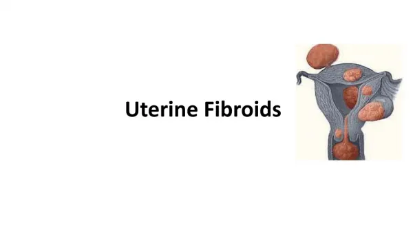
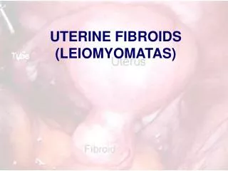
UTERINE FIBROIDS (LEIOMYOMATAS). What are they?. Smooth Muscle Tumor of the Uterus The most common uterine tumor Occurring in about 30% of women above the age of 30 years. Occurs up to 75% of hysterectomy specimens Symptomatic in 1/3 of cases. Patient Characteristics. Age: 30-40 years.

E N D
What are they? • Smooth Muscle Tumor of the Uterus • The most common uterine tumor • Occurring in about 30% of women above the age of 30 years. • Occurs up to 75% of hysterectomy specimens • Symptomatic in 1/3 of cases
Patient Characteristics • Age: • 30-40 years. • Rare before 30 or after 40 years • Parity: • Common in nulliparas, patients with low parity. • It is rare in multiparas. • Race: • 3-9 times more common in negroids. • Family history: • Usually positive. • Hyper-estrenemia: • Estrogen receptors (ER) more than the surrounding myometrium but less than those in the endometrium • Common in low parity. • Atrophies and shrinks after menopause. • Common association with other hyper-estrenic conditions as endometriosis, endometrial hyperplasia and endometrial carcinoma.
[60%] [20%] [15%]
Characteristics • Size • from microscopic to very huge size filling the whole abdominal cavity (up to 40 kg was recorded). • Shape • Spherical, flattened, or pointed according to the type. • Cut section: • On cut section,, whorly in appearance, and more pale than the surrounding uterine muscle. • Consistency: • firmer than the surrounding myometrium. • Soft fibroid occurs in pregnancy, cystic changes, vascular, inflammatory, and malignant changes. • Hard fibroid occurs in calcification. • Capsule: • Is a pseudo-capsule formed by compressed normal surrounding muscle fibres. • the blood supply comes through it, • it is the plain of cleavage during myomectomy • its presence differentiate the myoma from adenomyosis. • Blood supply: • Nourishes the myoma from the periphery, • The tumor itself is relatively avascular.
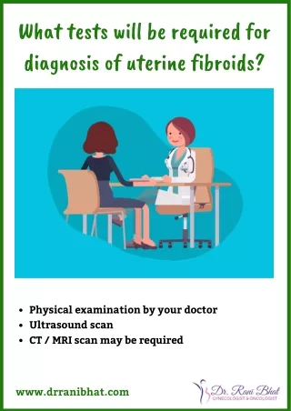
Presentations • Asymptomatic: • Accidentally discovered during examination. • It is the commonest presentation, especially in subserous and interstitial fibroids. • Vaginal bleeding: It is the commonest symptom, • Menorrhagia or polymenorrhea: (commonest): This occurs due to: • Associated hormonal imbalance and endometrial hyperplasia. • Surface ulceration of submucous fibroid. • Interstitial fibroid acts as F.B. preventing full contraction of myometrium to decrease blood loss. • Pelvic congestion. • Increased uterine size, vascularity, and endometrial surface area. • Metrorrhagia: due to: • In submucous fibroid due to ulceration of the surface, necrosis of the tip, or secondary infection. • Associated endometrial polyp. • Associated malignancy (cancer body or sarcomatous change). • Contact bleeding: (rare) • ulcerated or infected tip of submucous fibroid polyp. • Post-menopausal bleeding: • Either due to sarcomatous change or associated endometrial carcinoma. • Picture of iron deficiency anemia.
Presentations • Discharge: • Leucorrhea and mucoid discharge due to pelvic congestion. • Muco-sanguinous discharge with ulcerated fibroid polyp. • Muco-purulent discharge due to secondary infection. • Swelling: • Either abdominal swelling due to large fibroid or vaginal swelling due to a polyp. • Infertility [in 5-10% of cases]: • Most important is the underlying predisposing factor as anovulation and hormonal disturbance. • Broad ligamentary fibroid may stretch or distort the tubes. • Corneal fibroids may obstruct the uterine end of the tube. • S.M.F. acts as F.B. interfering with implantation. • Cervical fibroid may obstruct the cervical canal. • Associated endometriosis or endometrial hyperplasia. • Pain: uncommon • Intermittent colicky pain in submucous fibroid (acts as F.B. in the uterine cavity). • Dull-aching pain and congestive dysmenorrhea due to pelvic congestion. • Acute abdomen in red degeneration, torsion, ruptured vessel, and inflammation.
Presentations • Pressure symptoms • Cervical fibroid: • Anteriorly on the urethra causing acute retention of urine, or the bladder causing frequency of micturition. • Laterally on the ureters causing colic and back pressure on the kidneys. • Posteriorly on the rectum causing dyskasia, constipation, and sense of incomplete defecation. • Huge fibroid: • On the pelvic veins causing edema, pain, and varicose veins in the lower limbs. • On the GIT causing distension and dyspepsia. • On the diaphragm causing dyspnea. • Spontaneous abortion: • Before myomectomy [ 40%] • 20% after myomectomy.
Signs of fibroid • General examination: • signs of chronic anemia. • Abdominal examination: • large pelvi-abdominal swelling in huge fibroids. • Pelvic examination: • symmetrically or asymmetrically enlarged uterus. • Speculum examination • fibroid polyp.
Differential Diagnosis • Causes of symmetrically enlarged uterus: • Pregnancy • Subinvolution of the uterus. • Submucous or interstitial fibroid. • Metropathia hemorrhagica. • Adenomyosis uteri. • Carcinoma or sarcoma of the uterus. • Pyo, hemato, or physometra. • Causes of asymmetrically enlarged uterus: • Subserous fibroid. • Localized adenomyosis. • Ovarian, tubal, or broad ligamentary swelling. • Pregnancy in a rudimentary horn.
Management • Conservative Management • small asymptomatic fibroid, • fibroid in pregnancy or puerperium. • Just keep observation every 6 months. • Beware of underlying and/or associated pathology
Medical Treatment: • Pre-operative till the time of surgery. • Patient near the menopause, or newly married with minimal symptoms. • Red degeneration with pregnancy. • Lines of treatment: • Symptomatic: • Correction of anemia, • haemostatics, • analgesics, and anti-spasmodics (anti-PG). • Anti-estrogens: • large dose of progesterone, • Tamoxifen, Danazol, • LH-RH analogues • useful in decreasing the size and vascularity of the tumor by 50% which is beneficial before myomectomy
Surgical Management Myomectomy vs. Hysterectomy ??!! • Indications: • Symptomatic cases or uterus larger than 12 weeks size. • Suspected malignancy (rapidly enlarging or post-menopausal growth). • Multiple huge fibroids liable to complications. • Infertility.
Myomectomy • Abdominal Myomectomy • Vaginal Myomectomy • Endoscopic Myomectomy • Hysteroscopic • Laparoscopic • Embolization techniques ( Interventional Radiology)
Principle • Myomectomy aims at • removal of all the myomas, • with conservation of a functioning uterus to preserve the reproductive function. • Generally the morbidity is higher than those with hysterectomy. • It is associated with much blood loss • Liability of recurrence of fibroid. • Myomectomy is better reserved only for those keen to preserve the reproductive function.
Principle • The patient must be prepared for the possible need for an emergency hysterectomy. • Precautions to minimize blood loss during myomectomy: • The timing of operation is post-menstrual (minimal pelvic congestion). • Pre-operative LH-RH analogues: may be given for 3 months before surgery to reduce the size and vascularity of the myomas. • Intraoperative hemostasis • Vertical midline incision is the least vascular • application of Bonney’s myomectomy clamp or a rubber tourniquet • Use ring forceps to occlude the ovarian vessels • Careful dissection to enucleate all the masses is needed to avoid recurrence. • Avoid anesthetic agents that induce uterine relaxation (e.g. halothane). • Vasopressin (pitressin) 20 IU in 20 ml in normal saline are injected in the uterine wall at the site of incision. • Obliteration of the tumour cavities. • Buried sutures to the tumor bed after shelling out of the masses. • Use absorbable sutures. • Blood needs to be prepared for possible transfusion
Technique of abdominal myomectomy: • Preliminary diagnostic curettage to exclude endometrial carcinoma. • The uterine incision: • Avoid incisions on the posterior uterine wall, for the risk of adhesions to the bowel. • The smallest incision is designed to enable removal of as many lesions as possible. • Tunneling in the uterine wall is utilized to minimize many incisions and peritoneal trauma. • Try to avoid opening the endometrial cavity. • To keep the uterus anteverted • ventrosuspension or plication of the round ligaments and uterosacral ligaments. • Dextran solution, Ringer lactate solution or dexamethazone could be instilled in the peritoneal cavity to minimize postoperative adhesions.
Vaginal Procedures • Vaginal myomectomy: • Indicated when a fibroid polyp is not larger than 8 weeks pregnancy size. • The polyp is grasped and twisted until the pedicle tears. • If the pedicle is too thick it is cut with scissors. • A large polyp could be cut as piece-meal fashion (morcellation).
Hysterectomy • Patient around 40 years, and completed her family. • The number or site contraindicate myomectomy • Severe bleeding during myomectomy. • Major damage of the uterus by myomectomy which affects its function for pregnancy. • Recurrent fibroids. • Suspicious of malignancy
Secondary Changes in Fibroids • Degenerative • Vascular • Inflammatory • Malignant Changes
Degenerative Changes • Hyaline degeneration: • Commonest secondary change. • Usually starts around the menopause, and in the center of the fibroid. • Macroscopically, fibroid looks homogenous, waxy, soft, with loss of whorly appearance. • Fatty changes: • Likely to start around the age of menopause. • Lipids reach the fibroid through the blood, so fatty change starts at the periphery of the fibroid, resulting in a yellow soft fibroid.
Calcification: • Step following fatty change when fatty acids undergo saponification with Ca salts giving Ca stearate and palmitate, forming layers of calcifications. • Clinically, the fibroid become hard like bone (Womb stone). • Radiologically, show a radio-opaque shadow with typical onion skin appearance. • Red degeneration (Necrobiosis): • Usually occurs in the middle trimester of pregnancy, due to increased vascularity and venous stasis, the tumor enlarges with hemorrhage inside the tumor. • It is called necrobiosis because it shows dead parts (central) and living parts (peripheral).
Atrophic changes: • Atrophy occurs due to estrogen withdrawal as after menopause, puerperium, or anti-estrogen use. • All myomas decrease in size after the menopause except in calcification it remains stationary, or with malignant change or HRT it increases in size. • Myxomatous change: • Occurs near the menopause, in the center of the myoma, forming a gelatinous mucoid material which may undergo pseudo-cystic changes. • Pseudo-cystic changes: • A step following hyaline or myxomatous changes, when it liquefies & becomes soft in consistency.
Vascular Changes • Torsion (Axial rotation): • Occurs in moderate-sized, pedunculated, subserous fibroid with no adhesions. • The precipitating factor is sudden twisting movement as trauma, intestinal movement, or fetal kick, leading to axial rotation which is prevented from re-twisting by the lashing effect of the pulsating pedicle. • The clinical effects depend on the onset of torsion: • Sudden torsion leads to acute abdomen and necrosis of the tumor. • Gradual torsion leads to gradual decrease of the blood supply from the pedicle which ends in the development of parasitic tumor. • Telangeactasis: • Likely to occur with pregnancy, malignant change, and cervical fibroid due to increased vascularity. • There are numerous dilated blood vessels on the surface of the fibroid which may rupture leading to acute abdomen and internal hemorrhage. • Lymphangeactasis: • Likely to occur around the age of menopause as the fibroid is full of lymphatics. • Dilated lymphatic vessels on the surface may rupture leading to lymphatic exudates and strong adhesions. • Congestion and edema: A result of impaction, incarceration, torsion, infection, or pregnancy
Inflammatory changes • Ways of infection: • Trauma of submucous fibroid e.g. D & C or labor. • Near by inflammation e.g. appendicitis. • Blood-borne (very rare). • Result of infection: • The fibroid becomes congested, tender, and even abscess formation; it becomes soft and heals by adhesions to the surrounding
Malignant changes • Rare (0.5%) into leiomyosarcoma (round, spindle, mixed or giant cell histopathology types). • Symptoms suggestive: • The fibroid becomes more painful. • Post-menopausal bleeding or growth of the tumor. • Signs suggestive: • The fibroid become softer, tender, or fixed. • Rapid growth of the tumor.
Complications of fibroid • Degenerative changes. • Vascular changes. • Inflammatory changes. • Malignant changes. • Pregnancy complications e.g. abortion, and preterm labor. • Pressure complications on the urethra, bladder, ureters, rectum, and pelvic veins. • Rarely, chronic inversion of the uterus. • Polycythemia and hypertension due to the release of erythropoietic agent. • Infertility. • Secondary parasitic attachment of fibromyomas to other abdominal structures gaining another blood supply.
What is the effect of Fibroid on Pregnancy and Pregnancy on Fibroid?