Download

1 / 20
200 likes | 399 Vues
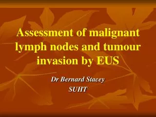
Assessment of malignant lymph nodes and tumour invasion by EUS. Dr Bernard Stacey SUHT. T1. T2. T3. T4. Malignant features of LNs. Size >1cm Hypoechoic Distinct margins Round shape All 4 present accuracy = 80% - All 4 features present only in 25%.

E N D
Assessment of malignant lymph nodes and tumour invasion by EUS Dr Bernard Stacey SUHT
T2 T3
Malignant features of LNs • Size >1cm • Hypoechoic • Distinct margins • Round shape • All 4 present accuracy = 80% - All 4 features present only in 25% Bhutani MS and Hawes R, Gastrointestinal Endoscopy 1997
Evidence for this? • Prospective study of 100 patients • All underwent resection • EUS LN features recorded wrt the 4 features • Correlation with histology • Sensitivity 89.1% • Specificity 91.7% • If LN imaged, likelihood of N1 disease = 86% • If no LN imaged 79% chance of N0 Catalano MF, Gastrointestinal Endoscopy 1994
More evidence for this • Prospective study of 457 patients • EUS features of LNs, cysts, wall lesions and extra-luminal lesions recorded • Correlation with clinical / histopathological data • EUS FNA for LNs • Sensitivity 92% • Specificity 93% • Complication rate 0.5% (higher for cystic lesions) Wiersema MJ, Gastroenterology 1997
Does this change outcome? • Retrospective study • 198 patients (SCC > adeno) • FNA in 20% • Sensitivity 97%, specificity 100% • This modified staging in 77.5% of cases • Surgery withheld from 60% of these Giovannini M, Endoscopy 1999
Changing outcome - Mayo clinic data • 74 patients - FNA on non-peritumoural LNs >5mm • half EUS + FNA, half EUS alone EUS aloneEUS + FNA Sensitivity 63% 93% * Specificity 81% 100% Complications 0 1
Is FNA the ideal test? • Availability • Complications • Interceding tumour • Sensitivity <100% (ie- false -ves) • Experience (US survey 2000 median number of EUS FNA performed / yr = 3) • LN characteristics may vary according to different primary tumour site
Intra-observer variability:agreement & reproducibility • Variability of T stage reporting even amongst experienced endosonographers • Best at extremes of T stage • Inexperienced --> poor T stage reporting but good at N stage • T2 least well reported • Repeatable in many studies • Pallazzo L, Hawes R Catalano MF, Gastrointestinal Endoscopy 1995
Computer assisted lymph node analysis • EUS images of LNs in oesophageal cancer correlated with histology after resection • EUS features of: • Echogenitcity • Whole-node heterogenicity • Regional variability • Assessed by computerized image analysis Loren DE et al, Gastrointestinal endoscopy 2002
Computer assisted lymph node analysis Benign v malignant • Hypoechoic p<0.04 • Heterogeneity p<0.004 • Regional variability p0.09 • Long/short axis p=0.05
Oesophageal cancer and PET • Metastases • Sensitivity 67% • Specificity 97% • Lymph nodes • Sensitivity 51% • Specificity 84% 16% false positives ie: benign - need FNA + ? Meta-analysis, Van Westreenen, J Clin Onc
T N M EUS CT PET Oesophageal cancer staging
The future • Prospective study into the Wessex experience: In what % of patients does EUS +/- FNA influence treatment in oesophageal cancer? Prospective / retrospective?