Mechanism of Smooth Muscle Contraction: The Sliding Filament Theory
The contraction of smooth muscle is primarily mediated by the sliding filament mechanism involving actin and myosin. In the resting state, actin is bound by caldesmon, while myosin light chain (MLC) is dephosphorylated, and intracellular calcium levels are low. A rise in intracellular calcium, triggered by neurotransmitters and hormonal signaling, initiates contraction. Calcium binds to calmodulin (Cam), activating myosin light chain kinase (MLCK) and inactivating caldesmon, allowing actin to be free for myosin binding. This leads to cross-bridge formation for muscle contraction.

Mechanism of Smooth Muscle Contraction: The Sliding Filament Theory
E N D
Presentation Transcript
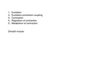
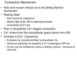
Contraction Mechanism • Actin and myosin interact via to the sliding filament mechanism • Resting State • Actin bound by caldesmon • Myosin light chain (MLC) dephosphorylated • Intracellular [Ca2+] low • Rise in intracellular Ca2+ triggers contraction • Ca2+ enters from the extracellular space (some from SR) • Increase in [Ca2+ ]caused by: • Excitation by neurotransmitters (extracellular Ca) • Hormonal signaling via receptors & 2nd messengers (SR Ca) • Ca flux can be inhibited by various inhibitory stimuli – hormonal & neuronal
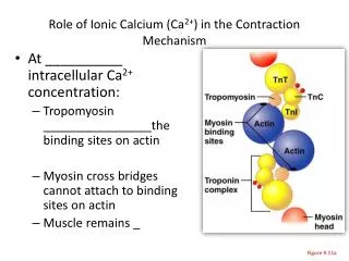
Role of Ca2+ for Contraction • Ca2+ binds to calmodulin (Cam) • Ca-Cam binds to & activates myosin light chain kinase (MLCK) • Ca-Cam binds to & inactivates caldesmon • Active caldesmon binds actin • Ca-Cam-inactivated caldesmon can’t bind actin • The free actin is available for myosin to bind • Activated MLCK phosphorylates myosin head region allowing cross bridges with actin to form