Download
1 / 39
500 likes | 881 Vues
Coral Bleaching and Coral Diseases: An Overview. J. Kilic. Phylum Cnidaria. Radial (or biradial) symmetry Diploblastic tissue organization Mesoglea between tissue layers Gastrovascular cavity Nerve net 2 body forms – polyp & medusa Cnidocytes (w/nematocysts). Phylum Cnidaria.

E N D
Phylum Cnidaria • Radial (or biradial) symmetry • Diploblastic tissue organization • Mesoglea between tissue layers • Gastrovascular cavity • Nerve net • 2 body forms – polyp & medusa • Cnidocytes (w/nematocysts)
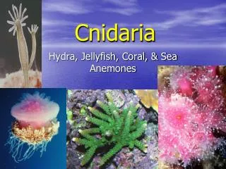
Phylum Cnidaria • 4 Classes • Hydrozoa - hydroids • Scyphozoa – true jellies • Cubozoa - Box jellies • Anthozoa – anemones & corals
Class Anthozoa • Corals & sea anemones • All Marine • Colonial (corals) or solitary (anemones) • No medusa stage • Polyps have a mesenteries and a pharynx leading to the GV cavity • Amoeboid cells in the mesoglea
Zooxanthellae • Algal symbionts • Most cnidarians possess the dinoflagellate Symbiodinium microadriaticum • Within the vacuoles of gastrodermal cells (about 50 dinoflagellates) • May contain as many as 30,000 symbionts per mm3 • The dinoflagellate enters the host in the egg or larval stage or the adult may engulf free algal cells.
Zooxanthellae • It is the pigments of the symbiotic algae that give corals their coloration • In most cases, the symbiosis is obligate • The host coral must live in shallow, clear waters (<75m) so the algae can photosynthesize. • Products of photosynthesis are translocated to the coral as carbon compounds. • The algae utilizes the coral’s nitrogenous wastes and acetate.
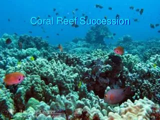
Coral Bleaching • Caused by the loss or large reduction in the zooxanthellea (or their pigment). • White calcium carbonate skeleton of the the coral becomes visible • Loss of zooxanthallea causes the corals to begin to starve. • Large number of environmental factors that may cause coral bleaching • Pollution, sedimentation, increased UV radiation, freshwater runoff, salinity changes, changes in atmospheric carbon dioxide • Strongest correlation has been found between sea surface temperatures (usually linked to ENSO) and bleaching
Coral Bleaching • In most species, temps above 32C along with increased UV radiation can trigger bleaching. • Although bleaching may be lethal, some corals do recover. • They may regain their symbionts when conditions return to normal (if timely) • During a bleaching event reproduction and growth are negatively affected and corals tend to be more susceptible to disease. • If conditions remain stressed for extended periods, death individual coral colonies or entire stretches of reef may occur.
Bleached section of The Great Barrier Reef off the coast of Queensland, AU
1998 Massive Bleaching Event • 1997-1998 experienced major bleaching events. • Every coral region in the world effected by bleaching in 1998 – the first global bleaching event • Triggered by severe ENSO conditions • Summer 1997-1998 at The Great Barrier Reef was the hottest on record • 67% inshore reefs showed “high or extreme” levels of bleaching (14% offshore) • Sea temps were 1-2C above long-term averages • On some reefs coral mortality reached up to 80%
More Recently… • 2002 ENSO conditions are thought to have triggered this major bleaching event. • 2005 NOAA reported a major bleaching event in the Caribbean. • Bleaching was reported from the entire area, the Florida Keys, Texas coast, Costa Rica, Tobago, Panama etc… • Bleaching coincided with areas that experienced levels of “high thermal stress”
“The DHW accumulates any HotSpots greater than 1 °C over a 12- week window, thus showing how stressful conditions have been for corals in the last three months. It is a cumulative measurement of the intensity and duration of thermal stress, and is expressed in the unit °C-weeks. DHWs over 4 °C-weeks have been shown to cause significant coral bleaching, and values over 8 °C-weeks can cause widespread bleaching and some mortality.” --NOAA Coral Reef Watch
Coral Disease • Viruses • Bacteria • Protozoan • Fungi • There are some diseases that appear to have no known pathogen associated with them
Rapid Wasting Disease • First observed in 1996 • Leaves skeleton exposed with no living tissue • Appears on the coral head first • Although the skeleton appears normal, when touched it simply crumbles • Cause is not yet confirmed, however • There has been observation of a filamentous fungus present on infected corals • Infected corals tend to be found where unfavorable algal species occur, particularly those that are often associated with excess nutrients from runoff and sewage.
Lethal Orange Disease Attacks the reef-building coralline algae Porolithon onkodes • Proceeds in an orange band leaving behind the white skeleton • Forms upright filaments and globules similar to slime molds • Coralline lethal disease is probably related but lacks the orange band • Believe to be a bacterial pathogen
Dark Spot Disease • Circular or irregular shaped dark spots appear on the surface of coral • Usually begins as purple or gray lesions • Sediment accumulates in the center of these patches • Cause is unknown, possibly a combination of pathogens
Coral Viruses (Vega, 2008) • Corals do not just have zooxanthellea as symbionts, they also have an array of microbial flora & fauna, much like we do • The “coral holobiont” refers to the coral, zooxanthellea & this normal flora & fauna. • Viruses present as a part of this normal state are often those that infect protozoans, metazoans, bacteria & archaea • Certain viruses and bacteria may be detrimental in times of stress • Temperature, nutrient levels, DOM
Herpes Viruses • Elevated abundance when temperature stress is applied • Herpes viruses tend to be under control as long as the coral is not stressed • Once stressed or compromised, the viruses become much more active • Positive correlation has been found between herpes genes and presence of coral tumors
Geminivirus • Single stranded DNA plant virus • Increased abundance with increased nutrients levels (ie fertilization runoff) • Symbiodinium abundance is negatively correlated with certain Geminiviruses • Zooxanthellea being reduced or lysed as a result of viral infection
Bdellovibrio Phages • Virus that infects bacterivorous bacteria • Increased numbers in the presence of increased DOM (carbon source) • Negative correlation between Bdellovibrio phages and heterotrophic bacteria • Suggests that Bdellovibrio phages kill the “good” bacteria that eat the “bad”
Coral Bacterial Infections • The good-guys: on 1 cm2 of coral, there may be 10 million bacteria and 1 billion archaea. • Many are part of the normal flora and are symbiotic • Control populations of harmful bacteria
White Band Disease I • Slow acting (1 cm/day) • Attacks Acroporid (branching) corals only • Tissue slowly peels off • White bands found at the base and middles of the coral • Gram negative rod shaped bacteria has been associated with the disease.
White Band Disease II • Fast acting (up to 10 cm/day) • Affects all corals Acroporid and non-Acroporid • Bleaching edge that precedes the dead egde • Bleaching edge may arrest and necrosis may catch up…if so, WBD I & II look very similar • Bacteria in the genus Vibrio have been found in the bleaching edge
Black Band Disease • Affects a large variety of corals • Slow acting • Black ring about a cm wide moving across the coral surface. • Leave behind bare skeleton • Caused by a number of bacteria resemblinga bacterial mat • Sulfur-reducers • Cyanobacteria
Red Band Disease • Host corals are limited to stag, star & brain corals • Brick red or dark brown microbial mat that advances across the surface of corals • Bacterial components of the microbial mat seem to differ from those found in black band disease
Black Aggressive Band Disease • Attacks a large variety of corals • Similar to BBD, but the band is much thinner • Actually a gray band • Cyanbacterium from the genus Spirolina is the most probable cause • Although others such as Ballesteros sp. have not been entirely ruled out
Yellow Band Disease • Yellow botch disease • Yellow pox disease • Distinctive yellow band that proceeds across the surface of the coral • Leaves behind a skeleton that is stained yellow (penetrates a few mm) • Bacterial pathogen is Vibrio sp.
Skeleton Eroding Band – A protozoan • Novel type of coral disease • Caused by Halofolliculina corallasia, eukaryotic protozoan • Damages not only the living tissue but also the skeleton of the coral. • Attacks a variety of corals • Colonies of black loricea (shields or houses) • When they reproduce asexually, they release chemicals toxic to the coral tissue.
Aspergillus – A fungus • Aspergillus is a ubiquitous genus of Ascomycetes soil fungi…in terrestrial ecosystems • Has been found in marine environments including coral reefs • Now known as the cause of brown sea fan disease • First observed in 1995 when a large percentage of purple sea fans appeared stuffed with material and were turning brown. • That material was fungal hyphae
Scolecobasidium – A fungus (Raghukumar 1991) • In the Bay of Bengal, 5 species of coral were regularly found with necrotic patches • Sections of the patches showed a dark brown hyphal network • Scolecobasidium a basidiomycete fungus was the causative agent • In contrast to most marine fungi identified to date being Ascomycetes
Citations • Raghukumar, Chandralata & Raghukumar, S. Fungal Invasion of Massive Corals. 1991 Marine Ecology 12 (3):251-260 • Kohlmeyer, B & Kohlmeyer, J. Mycological Research News, Letters: Fungi from Coral Reefs: A Commentary. 2003. Mycological Research 107 (4) 385-387 • Bruno, John F., Petes, Laura E., Harvell, C. Drew, Hettinger, Annaliese. Nutrient Enrichment can Increase the Severity of Coral Diseases. 2003. Ecology Letters 6: 1056-1061 • Vega Thurber, R., Barott, K., Rodriguez-Brito, B., Liu, H., Hall, D., Edwards, R.A., Desnues, C., Angly, F., Haynes, M., Wegley, L., and Rohwer, F. MetagenomicAnalysis Indicated that Stressors Induce Production of Herpes-like Viruses in the Coral Porites compressa. (in review, PNAS) • http://www.noaanews.noaa.gov/stories2005/s2526.htm • http://www.reef.crc.org.au/publications/brochures/1998event.htm • http://www.marinebiology.org/coralbleaching.htm • www.sbg.ac.at/ipk/avstudio/pierofun/aqaba/disease1.htm • http://www.livescience.com/environment/070620_microbes_corals.html