Download
1 / 47
570 likes | 2.24k Vues
Ocular Myasthenia Gravis: Past, Present, and Future. Victoria S. Pelak, MD Departments of Neurology and Ophthalmology University of Colorado Health Sciences Center. Ocular Myasthenia Gravis. 1. Definition and Natural History 2. Epidemiology 3. Anatomy & Pathophysiology

E N D
Ocular Myasthenia Gravis:Past, Present, and Future Victoria S. Pelak, MD Departments of Neurology and Ophthalmology University of Colorado Health Sciences Center
Ocular Myasthenia Gravis 1. Definition and Natural History 2. Epidemiology 3. Anatomy & Pathophysiology 4. Clinical features & Differential Dx 5. Diagnostic tests 6. Treatment 7. Future Options
Definition • Weakness and fatigability of cranial, limb, respiratory muscles • “generalized” • Levator palpebrae superioris, EOMs, and orbicularis oculi • “ocular” • 15% purely “ocular”
Natural History Ocular symptoms in Myasthenia Gravis: • 50% present solely with • 75-80% have on presentation • 90% eventually develop
Natural History(Grob et al. ’81) • ~2/3 will generalize • Who? • When? • first 7 months • OMG @ 1 year: 84% will NOT • OMG @ 2 years: 88% • OMG @ 3 years: 92%
Historical Perspective • Thomas Wills 1672 • Samuel Wilks 1877 • Ernst Sauerbrch 1912 • Mary Walker 1934 • C.E. Chang 1962 • 1970s Thomas Wills
Epidemiology: Incidence • Incidence MG: 4-14/100,000 age and gender related • generalized: early peak late peak • ocular: late peak
1915-34: 70% 1935-39: 40% 1940-57: 33% 1958-65: 14% 1966-85: 7% 1934: anticholinesterase 1939: assisted ventilation 1960: pressure or volume 1966: steroid use Epidemiology: Mortality (Grob et el. ’87)
Epidemiology: Associated Conditions • Thyroid dysfunction • Rheumatoid Arthritis • Ankylosing spondylitis
Anatomy & Pathophysiology • Anatomy • Neuromuscular junction • Pathophysiology • Causes • Autoimmune
Anatomy • Central nervous system • Peripheral nerve • Neuromuscular junction • Muscle • Combination
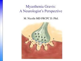
Electrical impulse Chemical impulse Electrical impulse Neuromuscular Junction
Neuromuscular Junction Disorders • Myasthenia Gravis • Lambert Eaton-Myasthenic Syndrome (LEMS) • Toxic or Metabolic • Botulism • Hypermagnesemia • Drugs (D-Penicillamine) • Organophosphate toxicity • Snake, spider, scorpion bites
Pathophysiology: Causes • Autoimmune • Neonatal • Congenital • Drug-induced
Neonatal Myasthenia Gravis • Passive transfer of IgG • 10 – 30% mothers with MG • 0 – 3 d after birth • Transient: 1-6 weeks • Weak cry, poor suck, hypotonia
Congenital Myasthenia Gravis • Genetic defects • Birth or infancy • Ocular +/- generalized • Fluctuate, stable
Drug-induced Myasthenia Gravis • D-Penicillamine
Autoimmune Myasthenia Gravis • Postsynaptic disorder • Decreased acetylcholine receptors • Immune-mediated
Clinical Features: OMG • Ptosis • Diplopia • Orbicularis oculi weakness
Ptosis • Isolation or with ophthalmoplegia • Fluctuates and shifts • Usually asymmetric • Examination: • Fatigability • “Cogan’s lid twitch” • Curtaining • Eyelid retraction
Ocular Motility Deficits • Any pattern • pseudo INO • pseudo 3rd, 4th, 6th • pseudo cavernous sinus syndrome • Exam changes • Medial rectus
Orbicularis Oculi Weakness • Common • Most commonly affected muscles: 1. levator palpebrae superioris 2. EOMs 3. orbicularis oculi 4. proximal limb 5. facial expression, mastication, speech 6. neck extensors
Ocular Myasthenia PEO Oculopharyngeal dystrophy Thyroid eye disease Intracranial mass lesion “Senile” ptosis Bulbar Dysfunction Motor neuron syndromes Oculopharyngeal dystrophy Polymyositis Generalized Myasthenia Lambert-Eaton syndrome Botulism Myopathy Differential Diagnosis
Diagnostic Tests • Anti-Acetylcholine Receptor Antibodies • Tensilon Test • Electromyography • Response to mestinon • Ice test
Anti-Acetylcholine Receptor Antibodies • Present in 80-90% of generalized • Present in 50% of ocular • No difference in severity, response, or prognosis
Tensilon Test • OMG: + 75% • False positive • Onset in 30s, lasts 1- 5 minutes • Heart disease and elderly • Atropine available
Electromyography • Repetitive nerve stimulation • 60-90% generalized • 20-30% OMG • Single fiber EMG • 90-100% generalized • 80-90% OMG
Mestinon Response • Poor in OMG • Ptosis • Motility
Ice Test • Ice pack on more ptotic lid x 2 minutes • Ptosis • 92% in MG • 0 non MG • Substitute for tensilon Borenstien et al. ‘75
Ice Test: Case of 75 year old woman • Negative antiacetylcholine receptor antibodies • Negative RNS and SFEMG • Negative Tensilon test x 2 • No response to mestinon • Ice pack at home when “eye” closed shut
Treatment • Cholinesterase inhibitors (Mestinon) • Immunosuppresion: • prednisone • cyclosporine • azathioprine (Imuran) • Thymectomy • Acute therapies • IVIg • Plasmapheresis
Cholinesterase Inhibitors (Mestinon) • Response often incomplete • ptosis • diplopia • Onset 30’, half life of 3-4 hours • SE: diarrhea • Caution: cardiac conduction defects
Prednisone • OMG: good response • Maintain high dose ~ 3 months or stable • Lowest effective dose • once determined alternate day therapy • majority need indefinitely • Caution: steroid-induced exacerbation
Cyclosporine and Azathioprine • Occasionally used in OMG • Toxicity • Indications: • resistant to steroids • need to reduce steroid dose • >50 mg qod • significant SE
Thymectomy • Definite indications: 1. Generalized: puberty – 60 years 2. Thymoma (15%) • OMG w/o thymoma: not rec • Response: months-years
Acute Therapies: IVIg and Plasmapheresis • Short term – transient (days to weeks) • Not indicated in OMG • Indications in GMG
Alternatives to Medical Treatment • Ptosis • ptosis crutches • ptosis surgery: not recommended • Diplopia • patch • prisms: too variable • strabismus surgery: poor outcome
Drug Precautions Antibiotics: aminoglycosides, neomycin, streptomycin, kanamycin, gentamicin, tobramycin, netilmicin, amikacin, Other: tetracycline, ciprofloxacin, erythromycin Anticonvulsants: dilantin Antimalarials: chloroquine, quinine Cardiovascular: quinidine, procainamide, verapamil, timolol, propanolol Ophthalmic: betaxolol, timolol Psychotropic: lithium, chlorpromazine Rheumatologic: D-penicillamine, chloroquine
Most Common Problems • Aminoglycosides • Beta blockers
Studies in OMG • Thyroid function tests • CT Chest • Review patient drug list • Tuberculin skin test • Rheumatologic screen
Future Options • Vaccine • Early immunosuppresion • injury to NMJ occurs during years 1-3 maximum weakness generalization prednisone treated OMG • trial: early IVIg
Future Options • Vaccine • Araga and Blalock ’94 • Anti-idiotypic antibodies • Prevention of experimental autoimmune myasthenia gravis
Future • Early immunosuppresion • injury to NMJ: year 1-3 maximum weakness generalization prednisone treated OMG • trial: early IVIg?