Download
1 / 41
420 likes | 633 Vues
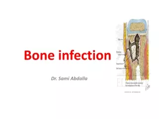
Specific bone and joint infection. 1- Tuberculosis. Incidence Showed a steady decline in its prevalence in developed countries during the 1960s and 1970s, due to the effectiveness of public health programs and advances in chemotherapy.

E N D
1- Tuberculosis Incidence Showed a steady decline in its prevalence in developed countries during the 1960s and 1970s, due to the effectiveness of public health programs and advances in chemotherapy. In the past two decades, however, the annual incidence has risen again which may be attributed to changes in population movements, the spread of intravenous drug abuse and the emergence of AIDS.
Predisposing factors • Chronic debilitating disorders, • Drug abuse, • Prolonged corticosteroid medication, • AIDS and other disorders resulting in reduced defense mechanisms.
Pathology • Mycobacterium tuberculosis ( usually human, sometimes bovine ) enters the body via the lung ( droplet infection ) or the gut ( swallowing infected milk products ) or, rarely through the skin. • Primary complex. The initial lesion in lung, pharynx or gut is a small one with lymphatic spread to the regional lymph nodes; this combination is the primary complex. Usually the bacilli are fixed in the nodes and no clinical illness results, but occasionally the response is excessive, with enlargement of glands in the neck or abdomen.
Secondary spread. If resistance to the original infection is low, widespread dissemination via the blood stream may occur, giving rise to military tuberculosis or meningitis. More often, blood spread occurs months or years later and bacilli are deposited in extrapulmonary tissues. Some of these foci develop into destructive lesions to which the term “ tertiary “ may be applied. • Tertiary lesion. Bones or joints are affected in about 5 ٪ of patients with tuberculosis. There is a predilection for the vertebral bodies and the large synovial joints.
The characteristic microscopic lesion is the tuberculous granuloma – a collection of epithelioid and multinucleated giant cells surrounding an area of necrosis, with round cells ( mainly lymphocytes ) around the periphery.
Bone lesions tend to spread quite rapidly. Epiphyseal cartilage is no barrier to invasion and soon the infection reaches the joint. Only in the vertebral bodies, and more rarely in the greater trochanter of the femur or small bones of the hands or feet, does the infection persist as a pure chronic osteomyelitis. • Caseation and infection may extend into the surrounding soft tissues to produce a cold abscess. This may burst through the skin, forming a sinus, or it may track along the tissue planes to point at some distant site. Secondary infection by pyogenic organisms is common.
Clinical features • There may be a history of previous infection or recent contact with tuberculosis. • The patient is usually a child or young adult. • Pain and (in a superficial joint) swelling. • In advanced cases there may be attacks of fever or lassitude and loss of weight. • Relatives tell of “ night cries “ . • Muscle wasting is characteristic. • Synovial thickening is often striking. • Movements are limited in all directions. • As articular erosion progresses the joint becomes stiff and deformed.
In tuberculosis of the spine, pain may be deceptively slight- often no more than an ache when the spine is jarred. Consequently the patient may not present until there is a visible abcess ( usually in the groin or the lumber region to one side of the midline ) or until collapse causes a localized kyphosis. Occasionally the presenting feature is weakness or loss of sensibility in the lower limbs.
X – ray • Soft - tissue swelling and periarticular osteoporosis are characteristic. • The bone ends take on a “ washed-out “ appearance and the articular space is narrowed. • Later on there is erosion of the subarticular bone, characteristically seen on both sides of the joint. • Cystic lesions may appear in the adjacent bone ends but there is little or no periosteal reaction. • In the spine: bone erosion and collapse around an intervertebral disc space; the soft tissue shadows may define a paravertebral abscess.
Investigations • The ESR: is increased. • CBC: relative lymphocytosis. • The mantoux or Heaf test: will be positive, these are sensitive but not specific tests. • If synovial fluid is aspirated, it may be cloudy, the protein concentration is increased and the white cell count is elevated. • Acid-fast bacilli are identified in synovial fluid in 10-20 ٪ of cases, and cultures are positive in over half. • A synovial biopsy is more reliable; sections will show the characteristic histological features, and acid fast bacilli may be identified; cultures are positive in over 80 ٪ of cases.
Diagnosis Features that should trigger more active investigation are: • A long history • Involvement of only one joint • Marked synovial thickening • Marked muscle wasting • Periarticular osteoporosis • A positive Mantoux test Synovial biopsy for histological examination and culture is often necessary.
Differential diagnosis: • Transient synovitis • Monoarticular rheumatoid arthritis • Subacute arthritis • Haemorrhagic arthritis • Pyogenic arthritis (in long standing cases).
Treatment Rest : This often involved splintage of the joint and traction to overcome muscle spasm and prevent collapse of the articular surfaces. With modern chemotherapy this is no longer mandatory; rest and splintage are varied according to the needs of the individual patient.
Chemotherapy : The most effective treatment is a combination of antituberculous drugs, which should always include rifampicin and isoniazid. A recommended regimen is rifampicin, isoniazide and ethambutol ( or pyrazinamide ) for 8 weeks, and thereafter rifampicin and isoniazide for a further 6-12 months.
Operation : Operative drainage or clearance of a tuberculous focus is seldom necessary nowadays. However, a cold abscess may need immediate draining. Once the condition is controlled and arthritis has completely subsided, normal activity can be resumed, though the patient must report any renewed symptoms. If , however, the joint is painful and the articular surface is destroyed, arthrodesis or replacement arthroplasty may be considered. The longer the period of inactivity, the less the risk of reactivation of the disease; there is always some risk and it is essential to give chemotherapy before and after the operation.
2- Brucellosis Brucellosis is an unusual but important cause of subacute or chronic granulomatous infection in bones and joints. • The organism : Brucella melitensis, brucella abortus ( from cattle ) and brucella suis ( from pigs ). • Mode of infection : Drinking unpasteurized milk or from coming into contact with infected meat. About 50 % 0f patients with chronic brucellosis develop arthritis.
Pathology: The organism enters the blood with infected milk products or, occasionally, directly through the skin or mucosal surfaces. It is taken up by the lymphatics and then carried by the blood stream to distant sites. Foci of infection may occur in bones (usually the vertebral bodies) or in the synovium of the larger joints. The characteristic lesion is a chronic inflammatory granuloma with round - cell infiltration and giant cells. There may be central necrosis and caseation leading to abscess formation and invasion of the surrounding tissues.
Clinical features: • Fever, headache and generalized weakness. • Followed by joint pains and backache. The initial illness may be acute and alarming; more often it begins insidiously and progresses until the symptoms localize in a single large joint ( usually the hip or knee ) or in the spine. The joint becomes painful, swollen and tender ; movements are restricted in all directions. If the spine is affected, there is usually local tenderness and back movements are restricted. The systemic illness follows a fluctuating course, with alternating periods of fever and apparent improvement ( hence the older term “ undulant fever “ ).
x-ray : • Loss of articular space, • Slowly progressive bone erosion and periarticular osteoporosis. • In the spine, there may bedestruction and collapse of adjacent vertebral bodies with obliteration of the disc.
Investigations: • Positive agglutination test ( titre above 1/80 is diagnostic ). • Joint aspiration or biopsy may allow the organism to be cultured and identified.
Treatment: Antibiotics; the infection usually responds to a combination of tetracycline and streptomycin for 3-4 weeks. Alternative drugs, which are equally effective and which may be used as combination therapy, are rifampicin and the newer cephalosporins.
Operation; an abscess will need drainage, and necrotic bone and cartilage should be meticulously excised. If the joint is destroyed, arthrodesis or arthroplasty may be necessary once the infection is completely controlled.
3- Spirochaetal infection Although rarely seen in many countries, spirochaetal bone infection is still quite common in some parts of the world.
Early congenital syphilis Treponema pallidum can cross the placental barrier and infect the fetus during the later half of pregnancy. However, bone changes do not usually appear until several weeks after birth. Clinical picture; The infant is sick and irritable. Hepatosplenomegaly. The first signs of skeletal involvement may be joint swelling and “ pseudoparalysis” – the child refuses to move a painful limb. Several sites may be involved, often symmetrically, with slight swelling and tenderness at the ends or along the shafts of the tubular bones.
x-rays: The characteristic changes are of two kinds: • Periostitis; diffuse periosteal new-bone formation along the diaphysis , usually of mild degree but sometimes producing “an onion-peel” appearance. • Metaphysitis; trabecular erosion in the juxtaepiphyseal region, showing first as a lucent band near the physis and later as frank bone destruction. • Serological tests are usually positive in both mother and child.
Late congenital and aquired syphilis Bone lesions in older childrin and adults are usually manifestation of tertiary disease, the result of gumma formation and endarteritis. Gummata appear either as discrete, punched-out radiolucent areas in the medulla or as more extensive destructive lesions in the cortex. The surrounding bone is thick and sclerotic. Sometimes the dense endosteal and periosteal new-bone formation is the predominant feature, affecting almost the entire bone (the classic “sabre tibia”).
Treatment • Antibiotics are ineffectual in tertiary syphilis. • An operation is occasionally needed if the gumma breaks down or if there is a pathological fracture.