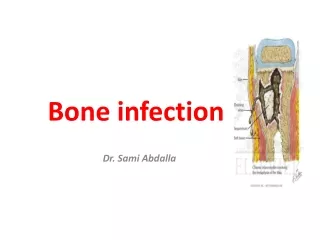
Bone Infection (osteomyelitis)
840 likes | 2.42k Vues
Bone Infection (osteomyelitis). ผศ.นพ.ยงศักดิ์ หวังรุ่งทรัพย์ ภาควิชาออร์โธปิดิกส์ จุฬาลงกรณ์มหาวิทยาลัย. Types of organism. Pyogenic osteomyelitis or arthritis Chronic granulomatous reaction Fungal infection Parasitic infestation. Route of Infection. Hematogenous system

Bone Infection (osteomyelitis)
E N D
Presentation Transcript
Bone Infection(osteomyelitis) ผศ.นพ.ยงศักดิ์ หวังรุ่งทรัพย์ ภาควิชาออร์โธปิดิกส์ จุฬาลงกรณ์มหาวิทยาลัย
Types of organism • Pyogenic osteomyelitis or arthritis • Chronic granulomatous reaction • Fungal infection • Parasitic infestation
Route of Infection Hematogenous system Direct invasion: Open Fx, operation, skin puncture Direct spreading
AcuteHematogenousOsteomyelitis • Common in children • Adult – lowered resistance by drug: immunosuppressive drug, debility disease: DM, AIDS - more common in vertebrae than long bone • Post-trauma: hematoma or fluid collection in bone
Pathogenesis Source of Infection Blood stream Metaphysis Venousstasis Bacterialcolonization
Etiology Aerobic organisms Gram positive : Staphylococcus aureus , Streptococcus pyogens Streptococcus pneumoniae -Gram negative : Haemophilus influenza, E.coli, Pseudomonas aeruginosa, Proteus mirabilis, Anaerobic organisms Bacteroides fragilis
Pathology Inflammation Suppuration Necrosis New bone formation Resolution
Inflammation • First 24 hours • Vascular congestion • Polymorphonuclear leukocyte infiltration • Exudation • Intraosseus pressure intense pain intravascular thrombosis ischemia
Suppuration • 2-3 days • Pus formation • Subperiosteal abscess via Volkmann canals • Pus spreading • epiphysis • joint • medullary cavity • soft tissue
Necrosis • Bone death by the end of a week • Bone destruction ← toxin ← ischemia • Epiphyseal plate injury • Sequestrum formation • small removed by macrophage,osteoclast. • large remained
New bone formation • By the end of 2nd week • Involucrum (new bone formation from deep layer of periosteum ) surround infected tissue. • If infection persist- pus discharge through sinus to skin surface Chronic osteomyelitis
Resolution Antibiotics Surgical drainage Infection is controlled Bone remodeling
Resolution • Infection is controlled • Intraosseous pressure release • With healing – new bone formation + periosteal reaction bone thickening and sclerosis • Remodeling to normal contour or deformity
Infection persist Chronic drainage Chronic Osteomyelitis
Signs and Symptoms in infant • Drowsy • Irritable • Fails to thrive • history of birth difficulties • History of umbilical artery catheterization • Metaphyseal tenderness and resistance to joint movement
Signs and Symptoms in child • Severe pain • Malaise • Fever • Toxemia • History of recent infection • Local inflammation pus escape from bone • Lymphadenopathy
Acute osteomyelitis in adult 1.Uncommon 2.History of DM. 3.Immunosuppressive drug 4.Drug addict 5.Elderly patients.
Signs and Symptoms in adult • Fever • Pain • Inflammation • Acute tenderness • Common site is thoraco-lumbar spine
Radiographic studies • มักจะเปลี่ยนแปลงหลังจากการติดเชื้อนานกว่า 10 วัน • เริ่มจาก rarefaction, area of lytic and sclerotic lesion, sequestrum and involucrum. • ควรเริ่มให้การรักษาทันทีก่อนจะเห็นการเปลี่ยนแปลงในภาพถ่าย X-ray
Bone Scan • 99m TC-HDP - sensitive - not specific • 67 Ga-citrate or 111 In-labeled leukocyte more specific
MRI ช่วยแยกpus กับbloodได้
Aspiration pus • confirm diagnosis • smear for cell and organism • culture and sensitivity test
Investigations • CBC • ESR • Hemoculture positive ~50% • Antistaphylococcal antibody titer (in doubtful case)
Differential diagnosis • Cellulitis • Acute suppurative arthritis • Acute rheumatism • Gaucher’s disease – Pseudo- osteitis, resembling osteomyelitis, enlargement of spleen and liver. Because of predisposing to infection, antibiotics should be given. • Sickle-cell crisis – mimic osteomyelitis, in endemic area of Salmonella, it is wise to treat with antibiotics until infection is excluded
Treatment for acute osteomyelitis • Supportive treatment • Splint • Antibiotic therapy • Surgical drainage
Supportive treatment • Analgesics • Correction of dehydration
Splint • Plaster slab • traction • Prevent joint contracture
Surgical drainage • Early treatment no need surgery • Late treatment surgical drainage about 1/3 of cases. If pus found and release no need to drill bone. But drilling one or two holes if no obvious abscess.
Antibiotics Initial antibiotics “ BEST GUESS ” - according to smear findings - according to incidences , age. Proper antibiotics - according to culture and sensitivities test
Acute osteomyelitis • When infection subside, movement is encourage. Walk with crutches and full weight bearing is possible after 3-4 weeks.
Complication • lethal outcome – rare • metastatic infection (multifocal infection) • suppurative arthritis • very young patient • metaphysis is intracapsular • metastatic infection
Complication • altered bone growth • chronic osteomyelitis - delay diagnosis and treatment - debilitated patients - compromised host
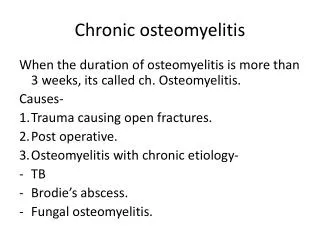
Chronic osteomyelitis • Sequel to acute hematogenous osteomyelitis • Usual organisms are staph. aureus, Escherichia coli, Strep. pyogens, Proteus and Pseudomonas (always mixed infections) • In the presence of foreign implants : Staph. Epidermidis is the commonest pathogen.
Pathology of chronic osteomyelitis • Bone is destroyed in a discrete area or diffuse • Cavities containing pus and sequestrum are surrounded by vascular bone and sclerosis bone resulted from reactive new bone formation • Sequestra, foreign implants act as substrates for bacterial adhesion, ensuring the persistence of infection and sinus drainage • Pathological fracture
Signs and Symptoms of chronic osteomyelitis • Pain • Pyrexia • Redness • Tenderness • Draining sinus • Excoriation of skin
Radiographic study • A patchy loss of bone density with thickening and sclerosis of the surrounding bone • Sequestra : dense fragment in contrast to surrounding vascularized bone • Sinogram may help to localize the site of infection
Radioisotope scanning • 99m TC-HDP Up take • 67 Ga-citrate or111In-labelled leukocyte more specific
CT – Scan and MRI • Show extent of bone destruction and reactive edema, hidden abscess and sequestrum • Pre-op planning investigation
Other Investigations • CBC • ESR • Antistayphylococcal antibody titers – Dx hidden infection and tracking progress to recovery • C/S from draining discharge R/O resistance bacteria