Benign and malignant Bone tumors
Benign and malignant Bone tumors. Bone tumors . Majority of Bone tumors are due to metastasis from other sites Most tumors have a predilection for a particular site in the body Causes: Idiopathic, bone infarcts (rare), chronic osteomyelitis , Paget disease, radiation, etc.


Benign and malignant Bone tumors
E N D
Presentation Transcript
Bone tumors • Majority of Bone tumors are due to metastasis from other sites • Most tumors have a predilection for a particular site in the body • Causes: Idiopathic, bone infarcts (rare), chronic osteomyelitis, Paget disease, radiation, etc.
Osteoma • Benign growths that involve the skull and facial bones • They may present singly or in multiples • Gardner syndrome is associated with facial osteomas ,multiple colonic adenomas and dermoid cysts • They may clinically present with a mass lesion that extends into the nasal cavity • There is no malignant association
Osteoidosteoma • Growth of this tumor involves the diaphysis of long bone • The lesions are usually less than 2cm. • There is cortical involvement • Signs/Symptoms: • Pain, characteristically more intense at night, relieved by NSAIDs and eliminated by excision • Vertebral lesions may cause scoliosis • Treatment: local excision
OsteoidOsteoma • Randomly oriented interconnected trabeculae of woven bone surrounded by a rim of osteoblas
Osteoblastoma • Similar to osteioidosteoma ; however it is larger than osteoidosteoma (>2cm) • Has a predilection for the vertebral column • Note: this tumor is not relieved by NSAIDs • Signs/Symptoms: • Pain • Gait disturbances • Treatment: local excision
Osteochondroma • These are relatively common slow growing, benign bone tumors • Probably not a true neoplasm, but rather a tumor produced by growth of aberrant foci of cartilage on the surface of bone • Autosomal dominant disorder of multiple herediatryexostosiswith risk of malignant transformation to chondrosarcoma
Enchondroma • These are benign tumors of hyaline cartilage that arise in the medulla (within the bone) • These are typically solitary lesions that arise in the metaphysealregion of the long bones • Enchondroma is the most common tumor of the bones of the hand
Enchondroma • 2 syndromes characterized by multiple enchondromas: • Ollier’s disease • Multiple enchondromas, usually unilateral • Maffucci’s syndrome • Multiple enchondromas associated with soft tissue hemangiomas , ovarian carcinoma and brain gliomas • Both disorders have 25% risk of malignant transformation to chondrosarcoma;
Enchondroma • Signs/Symptoms: • Usually asymptomatic lesions; pain with pathologic fracture • X-Rays shows O Ring - sign
Giant cell tumor • This is also known as osteoclastoma • It is relatively benign and uncommon; however, it is very aggressive • A majority of these tumors arise from the epiphyses of long bones • It is an expanding lytic lesion surrounded by a thin rim of bone
Giant cell tumor • They are usually present around the knee • The tumors are large and red-brown with frequent cystic degeneration • Signs/Symptoms: • Pain, loss of mobility, fracture • Treatment: curettage – surgery
Fibrous Dysplasia • McCune Albright syndrome • Jaffe syndrome • Monostotic • Ground glass radiographic appearance
Fibrous Dysplasia • Benign tumor • Localized developmental arrest; • All of the components of normal bone are present, but they do not differentiate into their mature structures. • Three clinical patterns: • Involvement of a single bone (monostotic); • Involvement of multiple bones (polyostotic); • Mccune-Albright syndrome -Polyostoticdisease, associated with café-au-lait skin pigmentations and endocrine abnormalities, especially precocious puberty.
Fibrous Dysplasia • Somatic gain-of-function mutation occurring during embryogenesis in the GNASgene • The mutation leads to-the production of a hyperactive guanyl nucleotide binding protein, encoded by the GNAS gene, that drives abnormal growth
Fibrous Dysplasia • Morphology: Well circumscribed intramedullary lesions that sometimes distort the bone. • Bone lesions are usually unilateral.skin lesions are seen on the same sides and affects ;The neck,back ,chest, shoulder and pelvis. • Micro :Curvilinear trabeculae of wooven bone that characteristically resembles Chinese letters. Osteoblastic components are mild and there may be cellular proliferation of fibroblasts.
Chondrosarcoma • These tumors can produce neoplastic cartilage • The tumor is made of atypical chondrocytes and chondroblasts with multiple nuclei • Pelvis, Shoulder and ribs • Signs/Symptoms: • Local swelling and pain in the enlarging mass • Treatment : wide local excision ; if aggressive chemotherapy is added
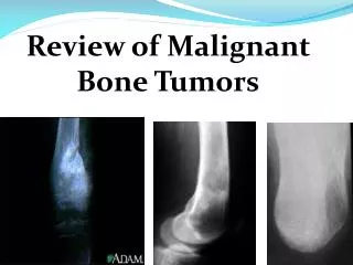
Osteosarcoma • This is also known as osteogenic sarcoma • This is the most common primary malignant tumor of bone • These tumors mainly arise from the metaphysesof the long bones, especially around the knee • Genetic mutations of the RB gene, p53, cyclin dependant cyclases .
Osteosarcoma • Malignant cells, produce osteoid • Most tumors arise de novo, though others arise in the setting of: • Paget’s disease • Previous radiation • Previous chemo (especially alkylating agents) • Chronic osteomyelitis
Osteosarcoma • The tumor frequently breaks through the cortex and lifts the periosteum, resulting in reactive periosteal bone formation • Secondary sarcoma occurs in elderly patients, especially associated with Paget disease, radiation,and chronic osteomyelitis these are highly aggressive and unresponsive to treatment
Osteosarcoma • Bilateral soft tissue masses, periosteal and cortical infiltration.
Osteosarcoma • Signs/Symptoms: • Pain and swelling in the enlarging mass • Pathologic fracture may be the first presenting symptom • X-Ray:A triangular shadow on x-ray between the cortex and raised periosteum is known asCodman’s triangle • Treatment: chemotherapy and surgery
Osteosarcoma • Mitotic figures lace like pattern of neoplastic bone.
Ewing’s sarcoma • Ewing sarcoma and primitive neuroectodermal tumors (PNETs) are primary malignant small round-cell tumors of bone and soft tissue • M>F,age 10-15 years. • Diaphysis of long bones • These two tumors only differ in the degree of differentiation Ewing’s sarcoma is undifferentiated and PNET is differentiated
Ewing’s sarcoma • Genetics plays a role patients with Ewing tumor several transocations.t (11;22) (q24;q12) is one of the most frequent. • M/E: the tumor is composed of sheets of uniform small, round cells ;Arrangement of tumour cell around a central fibrillary space is known as Homer Wright pseudorosettes
Ewing’s sarcoma • Signs/Symptoms: • May simulate osteomyelitis as patients often present with pain, fever and leukocytosis • Xrays may show periosteal reaction “onion skin” appearance • Treatment: chemotherapy , surgery, and radiation