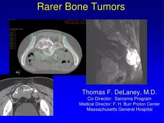
Benign bone tumors
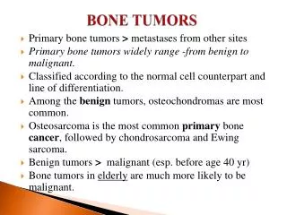
Benign bone tumors. DR: Gehan mohamed. Benign bone tumors. Osteoma osteoid osteoma giant osteoid osteoma (osteoblastoma) osteochondroma. Definition of osteoma:. A benign tumor composed of bony tissue, often developing from the skull. Osteoma in the frontal sinus.

Benign bone tumors
E N D
Presentation Transcript
Benign bone tumors DR: Gehan mohamed
Benign bone tumors • Osteoma • osteoid osteoma • giant osteoid osteoma (osteoblastoma) • osteochondroma
Definition of osteoma: • A benign tumor composed of bony tissue, often developing from the skull.
Osteoid Osteoma • An osteoid osteoma is a benign (non-cancerous), small tumor that usually grows in the long bones of a person. • Cell of origin : osteoblast. • Signs/Symptoms: • Pain specially at night relieved by NSAID and eliminated by excision. • Age: • 10-30 years • Sex: • M > F (2:1) • Anatomic Distribution: • It arises in the Metaphysis of bones • Over 50% of cases in femur or tibia • Nearly every location, most frequent in femur, tibia, humerus, bones of hands and feet, vertebrae and fibula
Osteoid osteoma :Central radiolucent nidus surrounded by thickened sclerotic bone
Osteoid osteoma :Central hemorrhagic nidus surrounded by dense rim of sclerotic bone
Histopathologic picture of osteoid osteoma: • It is formed of : - osteoid which is the unmineralized, organic portion of the bone matrix that forms prior to the maturation of bone tissue. - irregular bone trabecula
Histologic features of osteoid osteoma: the nidus of an osteoid osteoma demonstrates irregular masses of eosinophilic osteoid matrix (white arrow) and intensely stained bone trabeculae (black arrow) rimmed by osteoblasts (arrowhead).
Osteoid Osteoma • Prognosis/Treatment: • Surgical excision is treatment of choice • Recurrence can occur with incomplete excision of the tumor.
Osteoblastoma (Giant Osteoid Osteoma) • It is similar with osteoid osteoma in histology • But osteoblastoma differ in : Anatomic Distribution: - Predilection for vertebral column than long bones so Tumors of the spine can cause scoliosis and neurological symptoms. The lesion may clinically present with myleopathic and/or radicular symptoms. - much larger size (more than 2 cm up to 11.0 cm)
Exostosis (Osteochondroma) • Is a benign tumor . • Also known as exostosis • Children and teenagers most affected, Male >> female. • Clinically appear as slowly growing masses, painful if press on nerve tissue • Solitary or multiple • Multiple Hereditary Exostosis autosomal dominant disease with inactivation of both copies of EXT gene in growth plate chondrocytes
Osteochondroma • Benign projection of bone with cartilaginous cap • Occurs in epiphyseal plate and grows laterally • Exhibits cortex and medullary portion • Conversion to sarcoma rare (<1%) but higher in patients with hereditary syndrome. • May convert to malignancy if cartilage cap becomes thicker and contains disorganized calcifications
Osteochondroma • Develops in bones of endochondral origin and arises from the metaphysis near the growth plate of long tubular bones. • Occasionally develops from bones of the pelvis, scapula and ribs. • Ultrasound safe and inexpensive way to evaluate thickness of cartilaginous capsule • MRI method of choice to evaluate thicknesss of cartilaginous cap to rule out malignant conversion
- Radiograph can demonstrate that cortex of osteochondroma blends with cortex of normal bone
Osteochondroma:it consists of : -Fibrocartilagenous cap on top -Newly made bone trabecula Forms inner portion Medullary cavity of osteochondroma and bone are continuous