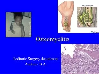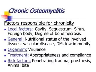
Chronic osteomyelitis
Chronic osteomyelitis. When the duration of osteomyelitis is more than 3 weeks, its called ch. Osteomyelitis. Causes- Trauma causing open fractures. Post operative. Osteomyelitis with chronic etiology- TB Brodie’s abscess. Fungal osteomyelitis. Pathology.

Chronic osteomyelitis
E N D
Presentation Transcript
Chronic osteomyelitis When the duration of osteomyelitis is more than 3 weeks, its called ch. Osteomyelitis. Causes- • Trauma causing open fractures. • Post operative. • Osteomyelitis with chronic etiology- • TB • Brodie’s abscess. • Fungal osteomyelitis.
Pathology. • Necrosis. stage of new bone formation involucrum. with sequestrum inside, there will always be a persistent discharging sinus. pus from bone escapes through multiple hole in involucrum
Clinical features- Pain, swelling. Discharging sinus. Bone thickening. Deformity. Joint stiffness. Shortening of limb, Pathological fracture. Sinus track malignancy. . • Discharging sinus
Investigation. • CBC, ESR. • Pus for c/s. • X – ray – sequestrum deformity. - periosteal thickening, sclerotic lesion. - irregular soft tissue shadow. - regional osteoporosis. - pathological fracture , micro fracture. • CT. • MRI
D/D- • TB osteomyelitis- watery discharge. - previous h/o TB, sinus with undermined margin with blue colour. 2. Ewing's sarcoma- A primary malignant tumor of bone, usually arising as a central tumor in long bone. (biopsy) 3. Soft tissue chronic infection. (X-ray)
Treatment. Supportive treatment . • Antibiotics – to prevent spread. • Surgery – sequestretomy + saucerization (excavation of the tissue of a wound to form a shallo )
Complication. • Joint stiffness. • Shortening. • Muscle contracture. • Pathological fracture. • Sinus track malignancy. • Amyloidosis.
Brodie’s abscess - it is an intraosseous abscess walled by reactive bone Cause- staph.aureus. Common site- metaphysis of long bone, cancellous bone. Clinical features- pain, fever, swelling, thickening of bone,
X- ray radiolucent area in sub periosteum surrounded by sclerotic bone. Treatment- Fenestration + antibiotics (two days of intravenous antibiotics and then oral antibiotics for six weeks )
Septic arthritis • Collection of pus inside the joint. • Common in knee and hip. • Cause- staph. Aureus, strept. pyogens, E. coli, pseudomonas, haemophilous influenza,
Mode of infection- • Haematogenous. • Direct penetrating wound to joint, • Joint aspirations, steroid injection, contrast injection, arthroscopy. • Spreading infection from near by bone.
Mode of infection Microbes synovial membrane inflammation (synovitis). increased synovium formation seorus/ seropurulent discharge erosion of cartilage - spreading of pus to the bone and destruction of bone. - escaping of pus outside the joint through sinus.
Clinical features • Neonates- increased pulse rate, fever, irritable, ignore feeding, swelling of joint, resist the movement of affected joint, pain. • Children- swelling, increased pulse rate, fever, restriction of movement of joint. • Adult- usually in immunocompromised or with other existing diseases.
Investigation. • CBC, ESR. • Pus for c/s , gram staining. • Blood for c/s • X-ray – initially normal. - initially increased joint space. - later joint space decreases. - osteopenia. • USG- collection of pus inside the joint space.
D/D • Acute osteomyelitis. • Haemarthaesis.- post traumatic. - aspiration. • Acute rheumatism. • Haemophilic joint • Gout.
Treatment • Supportive measures- 2 wks of IV antibiotics followed by 4-6 wks of oral antibiotics. • Symptomatic.(pain, fever) • Splintage of affected limb. • Surgery – arthotomy and evacuation of pus.
Complications. • Dislocaton of joint. • Deformity around joint. • Bony ankylosis.