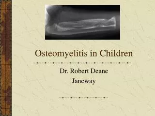
Chronic Osteomyelitis
Chronic Osteomyelitis. Factors responsible for chronicity Local factors: Cavity, Sequestrum, Sinus, Foreign body, Degree of bone necrosis General: Nutritional status of the involved tissues, vascular disease, DM, low immunity Organism: Virulence Treatment: Appropriateness and compliance

Chronic Osteomyelitis
E N D
Presentation Transcript
Chronic Osteomyelitis Factors responsible for chronicity • Local factors:Cavity, Sequestrum, Sinus, Foreign body, Degree of bone necrosis • General:Nutritional status of the involved tissues, vascular disease, DM, low immunity • Organism:Virulence • Treatment:Appropriateness and compliance • Risk factors:Penetrating trauma, prosthesis, Animal bite
Chronic Osteomyelitis Types • A complication of acute Osteomyelitis • Post traumatic • Post operative
Chronic Osteomyelitis Clinical picture • Continuous or intermittent suppuration and sinus formation with acute exacerbations. • Pain, fever, redness, and tenderness during acute exacerbations. • Discharging sinus with +ve/-ve culture. • Pathological fracture.
Chronic Osteomyelitis Investigation • Lab tests/ culture • Plain X-ray: Bone rarefaction surrounded by the dense sclerosis, sequestration and cavity formation • Sinogram • Bone scan & gallium scan To detect chronic multifocal osteomyelitis • CT Scan & MRI • Biopsy
Chronic Osteomyelitis Complications • Recurrence & Recurrence & Recurrence • Pathological fractures • Growth disturbance • Amyloid disease • Epidermoid carcinoma of the fistula
CHRONIC OSTEOMYELITIS • Sequel of acute/open fracture/opt. • C/F: pain, discharging sinus, scars • Xray- bone resorption, sequestra, • CT/MRI: extent of bone loss, oedema, hidden abscess • Lab-raised ESR pus-cs
TREATMENT • Antibiotics. • Local treatment. • Operations. • After care.
TUBERCULOSIS • A surgeon could gain experience in the management of TB. Of bone and joint only if he choose to work in econmically less developed countries. (edit, Br.Med.J>1968)
T.B. OSTEOMYELITIS • 1/3 population infected. • Over 80,000 people in Nepal have TB. • 22,000 develop Pul. TB every year. • Total 50,000. • 10,000 die of TB. • LEADING CAUSE OF DEATH. • 1 – 3% skeletal TB
TB • Cause by Mycobacterium tuberculosis, occasionally by M.bovis/africanum. • Also known as tubercle bacilli as they produce lesion – tubercles. • Acid fast bacilli. • Transmission – airborne droplets. • Risk- extent of exposure to droplets and susceptibility to infection.
TB Primary infection • Exposure to tubercle bacilli – Lungs – multiplication of bacilli in terminal alveoli (Ghon focus) – lymphatic drain it to hilar lymph nodes – (PRIMARY COMPLEX) – BLOOD – SPREAD.
TB • C/F:Cough >3wks.,sputum production, weight loss, monoarticular. • Respiratory – haemoptysis, chest pain, breathlesness. • Constitutional:fever/night sweat , tiredness , loss of appetite. • Physical sign: non specific,muscle wasting, loss of ROM
TB • 3 days sputum. • Ziehl-Neelsen stain. • X-ray: cavitation, infilteration, lymphadenopathy. • Full blood count:Relative lymphocytosis,^ ESR,Anemia. • Serology. • Lymphnode biopsy. • CT/ MRI
BONE TUBERCULOSIS • Spread from primary complex to any bone/joints. • Can effect any bone but the weight bearing bones are more likely to be affected. • Spine –commonest, hip, knee , foot.
STAGES OF ARTICULAR TB • 1 – SYNOVITIS. • 2 – EARLY ARTHRITIS. • 3 – ADVANCED ARTHRITIS. • 4 – ADV.ART. PATHOLOGICAL DISLOCATION / SUBLUXATION. • 5 – AFTER MATH TERMINAL OF GROSS ARTHRITIS.
Caseous exudative- more destruction, exudation & abscess formation. Symptoms more marked. Onset is less insidious. Granular type – less destructive. Abscess formation rare. Dry lesion. adults TB - TYPES
TUBERCULOSIS • Spine is the most common site of skeletal TB
TUBERCULOSIS • Pathology • Blood borne - settles in vertebral body anteriorly • usually more bone destruction, more sequestra, larger abscess, gaseous pus than pyogenic OM • intervertebral discs preserved until late disease
TB SPINE -Classification • 1- pre-destructive • 2- early destructive. • 3- mild angular kyphos. • 4- moderate angular kyphos. • 5- severe kyphos (humpback)
Predestructive –straightening of curvatures , spasm of perivertebral muscles, MRI-marrow oedema. Early destructive – Diminished disk space and paradiscal erosion.MRI-marrow oedema and break of osseous margin.CT-marginal erosion /cavitaion STAGES –1 & 2
TB SPINE –D/D • AGE- anomalies. • Infection. • Tumour. • Traumma. • Osteoporosis ,Osteochondrosis. • Spondylolisthesis.
TB SPINE • C/F:Back pain of variable duration, fever and weight loss. • O/E: local tenderness, spasm, mild kyphosis-late Gibbus, cold abscess and paraparesis. • DIAGNOSIS: XRAY-erosion of the anterior edges of the superior and inferior boarders of adjacent vertebral bodies with narrowing of disc space. • USG :paravertebral abscess. • Biopsy/ CT scan
TB HIP • C/F: pain/limping, irritable hip –child. Gradual loss of range of movement, flexion deformity, wasting of thigh muscles. • Xray: both hip to compare.Early changes –rarefaction of the bone and widening of the joint space, later destruction of the joint. • Synovial bioposy.
TB KNEE / ANKLE. • C/F: pain and synovial swelling, muscle wasting., contracture, draining sinuses. • X-ray. • Synovial biopsy.
PRINCIPLES OF MANAGEMENT OF TB • General. • Rest, mobilization & brace. • Abscess, effusion & sinuses. • Antitubercular drugs. • surgery
TB PROBLEMS • Diagnosis. • Treatment . • Anti tuberculous drugs.
Tuberculous lesion • Resolve completely. • Complete healing with varying degree of deformity / loss of function. • Lesion may be complete walled off and the caseous tissue may calcified. • Persist as a low grade ch.fibromatous granulating & caseating lesion. • Infection may spread. • Damage growth centre with shortening.
CONCLUSION • Slow progressive course of clinical symptoms and radiological signs of tuberculosis creates difficulty in early diagnosis. • Anti tuberculous treatment is effective but the functional outcome depends on early diagnosis before the development of radiological evidence of joint destruction. • Always keep TB in D?Diagnosis
Thank you for not sleeping Now you can ask your questions ???