. OSTEOMYELITIS
. OSTEOMYELITIS. DR SHAILESH. DEFINITION. Osteomyelitis is an inflammation of the bone caused by an infecting organism Osteon -bone Myelo -marrow The infection mainly involves marrow spaces, haversian canals, and subperiosteal space. Bone is involved secondarily. CLASSIFICATION.

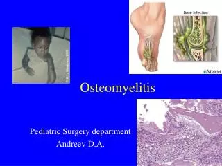
. OSTEOMYELITIS
E N D
Presentation Transcript
.OSTEOMYELITIS DR SHAILESH
DEFINITION • Osteomyelitis is an inflammation of the bone caused by an infecting organism Osteon-bone Myelo-marrow • The infection mainly involves marrow spaces, haversian canals, and subperiosteal space. • Bone is involved secondarily.
CLASSIFICATION • Based on duration of symptoms: Acute Sub acute Chronic - Active - Dormant
CLASSIFICATION • Based on mode of spread: a)Endogenous (Haematogenous) b)Exogenous • Based on host response: a)Specific b)Non-specific
It is a rapidlydestructive pyogenic infection, usually haematogenous inorigin, starting in metaphysisof an actively growing long bone • Seen in infants and children
It may occur in adults also due to spread of infection directly from another site or through compound fractures. • Haematogenous spread is rare
ETIOLOGY • Disease is common during the period of active bone growth (below 2 years and 8-12 years). • Sex - Male : Female - 4:1 • Site -Metaphysis, mainly in children (60-70% around knee).
RISK FACTORS • Poor nutrition and unhygienic surrounding. • Trauma (more in men and boys). • Skin, dental, respiratory, GI, UT infections. • Bacterial endocarditis. • Burns
RISK FACTORS • Leukopoenia and leukocyte dysfunction. • Iatrogenic intervention (catheters) • I.V. drug abuse • Sickle cell anaemia • Haemodialysis/immune suppressive therapy
BACTERIOLOGY Gram positive Staph. aureus 60-90% Streptococcus –multiple lesions Streptococcus epidermis, Enterococci, Clostridium, Bacillus, Diphteroids, Listeria.
BACTERIOLOGY Gram negative Pseudomonas (greenish discharge) E.coli, Proteus, Klebsiella, Haemophilus influenza-infants Salmonella, brucella, niesseria, bacterioids.
BACTERIOLOGY Specific type • Tuberculosis • M.leprae, • Spirochetes-T.pallidum • Actinomycosis,Candida,Cryptococcosis,Coccidiodomycosis • Viral-small pox • Parasitic
BACTERIOLOGY 50 45 40 35 30 Percent 25 20 15 10 5 0 CNS E-coli MRSA Staph epi Strep Gp B Strep viridans Staph aureus Pseudo aeruginosa IDLinks Speakers Club
Hair pin bend vessels in metaphysis • As there is increased vascularity to metaphysis causing pooling of blood, it has been aptly called ‘a lake of blood’. • Immature cells of metaphysis due to high cell turnover.
Relative lack of phagocytosis. • Presence of degenerating cartilage cells which acts as a good culture media. • Presence of end arteries in metaphysis. • More prone for trauma. • Presence of a single endothelial lining in metaphyseal arteries.
CLINICAL FEATURES • Antecedent infection is usually present /history of trauma. • Symptoms of Acute illness appears like irritability, restlessness,headache,vomiting, convulsions, chills, febrile with increase PR.
CLINICAL FEATURES • Pain • Initially no swelling is present but within few days, "the soft tissues becomes oedematous • Limb will be in a position of ease with restricted movements and surrounding muscle spasms.
CLINICAL FEATURES • Sympathetic synovitis of adjacent joints may be present. • If patient is not treated, he becomes apathetic and unconscious and continues fatal termination.
INVESTIGATION • Blood investigation • Aspiration of pus & blood culture • X-ray • Bone scan • MRI
DIFFERENTIAL DIAGNOSIS • Rheumatic fever • Ewing’s tumor • Acute suppurative arthritis
PRINCIPLES OF TREATMENTNADE(1983) • An appropriate antibiotic will be effective before pus formation. • Antibiotics will not sterilize avascular tissues and purulent material that must be removed surgically.
3. If such removal is effective, then antibiotics should prevent their reformation and therefore primary wound closure should be safe. 4. Surgery should not further damage already ischaemic bone and soft tissues. 5. Antibiotics should be continued after surgery.
CONSERVATIVE TREATMENT • The affected part should be positioned comfortably at rest. • General supportive measures. • If subperiosteal and bone aspiration do not show any abscess, IV antibiotics are started.
SURGICAL TREATMENT Indications for Surgery • Presence of an abscess requiring drainage. • Failure of patient to improve despite appropriate IV antibiotic treatment. Objective of surgery is to drain any abscess cavity and remove all dead and necrotic materials.
AFTER TREATMENT • Part should be protected for several weeks to prevent pathological fracture. • IV antibiotics should be continued post operatively for a short course followed by oral antibiotics.
COMPLICATIONS Early • Septic arthritis • Tenosynovitis • Thrombophlebitis • Death from associated septicaemia
COMPLICATIONS Late • Pathological fracture • Local growth disturbances : Over growth - due to prolonged hyperaemia. • Premature cessation - Due to epiphyseal plate destruction. • Deformity due to epiphyseal plate destruction.
PROGNOSIS • About 90% of cases diagnosed early and treated energetically may be expected to resolve completely. • The organism • The bone infected • The age of the patient
Differs from acute OM in the severity of symptoms and signs. This may be due to • Increased host resistance • Low bacterial virulence • Antibiotic therapy before symptoms occur.
CLASSIFICATION • Type-I :Metaphyseal lesion • Type-II:Eccentric metaphyseal lesion with cortical erosion • Type-III: Diaphyseal cortical lesion
CLASSIFICATION Type-IV :Lesion is a medullary abscess in diaphysis with periosteal reaction Type-V :Primary sub acute epiphyseal OM. Type-VI :Subacute OM crossing epiphyseal plate to involve both metaphysis and epiphysis.
CLINICAL FEATURES • Pain without constitutional symptoms. • ESR elevated in 50% of cases. • Blood culture negative. • Pus C/S positive in 60% patients.
DIAGNOSIS • On clinical suspicion and radiographic imaging. • Bacteriology : Staph, aureus and epidermidis common
TREATMENT • Typical lesions - Antibiotics for 6 weeks. • Diagnosis in doubt – Biopsy and curettage of lesion and antibiotics.
When acute OM was untreated/ inadequately treated or due to haematogenous spread of low virulence or infection through external wound.
CLASSIFICATION Cierny and Mader classification - based on anatomic and physiologic criteria. Anatomic Criteria: Intramedullary Superficial Local Diffuse with segmental bone loss.
CLASSIFICATION Physiologic Criteria: • Normal immune system and adequate soft tissue envelope. • Local /systemic /both compromised patients. • When the results of treatment are potentially more damaging than the presenting condition
SEQUESTRUM It is a microscopic or macroscopic fragment of necrotic, usually, cortical bone found at the nidus of the infection within the bone. MERCER