Download
1 / 15
150 likes | 380 Vues
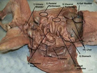
DIAGNOSIS OF BLADDER CALCULI. الدكتور:جاسم عبد الحسن اخصائي جراحة الكلى والمسالك البولية. FEMALE URINARY TRACT. MALE URINARY TRACT. EPIDEMIOLOGY. Bladder lithiasis is believed to make up 5% of all urinary calculi.

E N D
DIAGNOSIS OF BLADDER CALCULI الدكتور:جاسم عبد الحسن اخصائي جراحة الكلى والمسالك البولية
FEMALE URINARY TRACT MALE URINARY TRACT
EPIDEMIOLOGY Bladder lithiasis is believed to make up 5% of all urinary calculi. The majority of bladder calculi occur in adults, particularly in elderly men with bladder outlet obstruction. However, groups at risk for urinary stasis and infection are also at risk for bladder stone formation.
PATHOPHYSIOLOGY • Unlike the rapid clearance rates of urine in the renal tubules, the bladder is a reservoir for relatively stagnant urine and its precipitates. The presence of infection or poor bladder emptying may allow for the supersaturation and heterogeneous nucleation around a nidus. Subsequent aggregation results in crystal and stone formation.
CLASSIFICATION Vesical calculi can be classified as :- • migrant. • primary idiopathic, or • secondary calculi, which include calculi related to urinary stasis, infection, and foreign bodies.
Migrant Calculi • Migrant bladder calculi are formed in the upper tracts, pass into the bladder, and are retained there. Most calculi that migrate out of the ureter into the bladder are smaller than 1 cm and, in adults, are easily passed per urethra especially in female.
Primary Idiopathic (Endemic) Calculi • Endemic bladder stones form in children in the absence of obstruction, local disease, neurologic lesion, or known primary infection. it is rare in developed countries. However, endemic bladder calculi remain common in infants and children of lower socioeconomic background in North Africa and the Middle and Far East. Stone formation results from dietary and nutritional deficiencies.
Secondary Bladder Calculi • These secondary bladder calculi are most often related to urinary stasis or recurrent urinary tract infection due to bladder outlet obstruction or neurogenic bladder dysfunction. Patients with foreign bodies in the urinary tract are also at risk for development of calculi.
CLINICAL PRESENTATION • The clinical presentation of bladder calculi varies depending on stone size and location, as well as patient characteristics. • While they may be found incidentally in an asymptomatic patient during imaging for other reasons, calculi typically present with classic findings, which may include dysuria, frequency, nocturia, suprapubic pain, terminal gross hematuria, intermittency and urinary retention These symptoms may be relieved with a supine or head-down position, which may change the position of an obstructing stone at the bladder neck.
Urinalysis may reveal microscopic hematuria, pyuria, bacteriuria and crystalluria, with urine cultures demonstrating urea- splitting organisms.
Due to the inaccuracies of plain film radiographs, sonography and computerized tomography are the imaging studies of choice for the diagnosis of bladder calculi. • Cystoscopy can be performed to: -confirm radiographic findings. - help determine causative factors and, - plan for future operative intervention .
Plain film showing vesicle stone due to foriegn body(retained ureteric stent)