Chromosomes
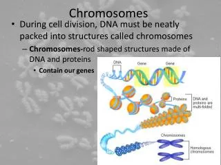
Chromosomes. Chromosome Structure. Prokaryotic chromosome. The term “prokaryote” means “primitive nucleus”. Cell in prokaryotes have no nucleus. The prokaryotic chromosome is dispersed within the cell and is not enclosed by a separate membrane.

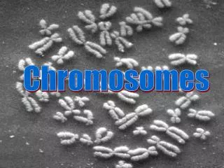
Chromosomes
E N D
Presentation Transcript
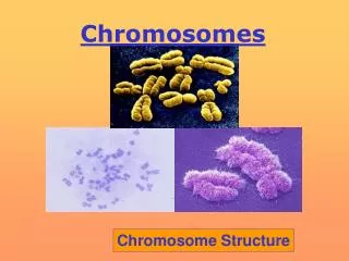
Chromosomes Chromosome Structure
Prokaryotic chromosome • The term “prokaryote” means “primitive nucleus”. Cell in prokaryotes have no nucleus. The prokaryotic chromosome is dispersed within the cell and is not enclosed by a separate membrane. • Much of the information about the structure of DNA has come from studies of prokaryotes, because they are less complex (genetically and biochemically) than eukaryotes. • Prokaryotes are monoploid = they have only one set of genes (one copy of the genome). • In most viruses and prokaryotes, the single set of genes is stored in a single chromosome (single molecule either RNA or DNA). • The smallest known RNA viruses have only three genes. • The smallest known DNA viruses have only 9 to 11 genes.
Prokaryotic chromosome • The bacterial chromosome must be tightly packed to fit into the small volume of the bacterial cell. The contour length of the circular DNA molecule present in the chromosome of the bacterium Escherichia coli is about 1500 µm. Because an E. coli cell has a diameter of only 1 to 2 µm, the large DNA molecule must exist in a highly condensed (folded or coiled) configuration. • Compacting the DNA involves supercoiling, or further twisting the twisted chromosome. • The chromosome's fifty or so DNA domains are held together by a scaffold of RNA and protein, and the entire nucleoid is attached to the cell membrane. • This membrane attachment aids in the segregation of the chromosomes after they replicate in preparation for cell division. • Bacteria lack the histone proteins that are found bound to DNA and that form a nucleosoms of eukaryotic chromosomes.
Prokaryotic chromosome • DNA molecule in an E. coli chromosome is organized into 50 – 100 domains or loops. • Replication of the circular chromosome begins at a single point, called OriC, and proceeds in both directions around circle, until the two replication forks meet up. • The results is two identical loops. Replication takes approximately forty minutes. • In 1997 F Blattner and colleagues published the sequence of 4,639,221 base pairs of the K-12 laboratory strain. E. coli is estimated to have 4,279 genes. • Many sets of genes on the E. coli chromosome are organized into operons. • An operon is a set of functionally related genes that are controlled by a single promoter and that are all transcribed at the same time. • It is also quite common for bacterial species to possess extrachromosomal genetic elements called plasmids. These are small, circular DNA molecules which, when present, vary in umber from one to about thirty identical copies per cell. • Plasmids include the fertility factor, as well as plasmids that carry drug-resistance genes.
Diagram of the structure of the functional state of the E. coli chromosome.
Eukaryotic chromosome • Eukaryotic genomes contain levels of complexity that are not encountered in prokaryotes. • Although eukaryotes have only about 2 to 15 times as many genes as E. coli, they have orders of magnitude more DNA. Moreover, much of this DNA does not contain genes, at least not genes encoding proteins or RNA molecules. • Eukaryotes enclose their genetic material in a specialized compartment called nucleus. • The basic component of the eukaryotic chromosome is its DNA, which contains all of the genetic material responsible for encoding a particular organism. • This DNA is packaged into several chromosomes, and each chromosome is present in two (diploids) or more (polyploids) copies.
Eukaryotic chromosome - organization • In human DNA has a total length of 1.8 meters and must fit into nucleus with an average diameter of 6 µm. • This feat is accomplished in part by the packaging of the DNA into chromatin, a condensed complex of DNA, histones, and nonhistone proteins. • The histones of all plants and animals consist of five classes of proteins. These five major histone types, called H1, H2a, H2b,H3, and H4, are present in almost all cell types. • The basic unit of chromatin is the nucleosome. The nucleosome is composed of approximately 146 base pairs of DNA wrapped in 1.8 helical turns around an eight-unit structure called histone protein octamer. • This histone octamer consists of two copies each of the histones H2a, H2b, H3, and H4. • The space in between individual nucleosomes is referred to as linker DNA, and can range in length from 8 to 114 base pairs, with 55 base pairs being the average. • Linker DNA interacts with the linker histone, called H1.
Chromatin and Chromosomes • Chromosomes linear structures in eukaryotic cells on which genes are arranged • Cells not in division genetic material is in an uncoiled state diffuse network Chromatin network or Chromatin • Each species chromosome number and structure • Distinguish a species somatic chromosome number
Heterochromatin vs. Euchromatin • Chromatin can be divided into two regions – euchromatin and heterochromatin, based on its state of condensation. • Most of the cellular chromatin is euchromatin, which has a relatively dispersed appearance in the nucleus. It condenses significantly only during mitosis. Genes within euchromatin can be transcriptionally active or repressed at a given point in time. • Heterochromatin is condensed in interphase, frequently is localized at the periphery of the nucleus, usually does not contain genes that are being expressed, and has late replication just prior to cell division. • Heterochromatin can be subdivided into constitutive and facultative heterochromatin. Constitutive heterochromatin is always inactive. Facultative heterochromatin refers to DNA sequences that are specifically inactivated as the result of development or regulatory event. • An example of facultative heterochromatin is the mammalian X chromosome.
Human metaphase chromosome showing the presence of 30-nm chromatin fibers. Each chromatid contains one large, highly coiled or folded 30-nm fiber.
Chromosome Characterization • Chromosomes linear with 2 telomeres and a constricted region called the centromere Centromere position • Metacentric : • Submetacentric : • Acrocentric : • Telocentric :
Chromosome Characterization cont. • Chromosomes usually occur in pairs homologous chromosomes gene sites called loci Heterozygous locus Homozygous locus • Homologous chromosomes not identical on a molecular level
Chromosome Number • Two types of chromosomes: Autosomes: all chromosomes other than sex chromosomesand occur in the same frequency in both sexes Sex chromosome: X and Y chromosomes that determine sex • Diploid organisms 2n = 2x n = gametic number haploid genome 2n = zygotic number diploid (2 x gametes) x = basic number (one set)
Chromosome Number cont. Variations in chromosome number • In humans x = 23 sets in mature gametes 2n = 46 diploid number in cells • More than one basic number: x-polymorphism in impala population x=29 and x=30 3 possible 2n numbers 2n=58; 59 and 60 • Hymenoptera (bees ands ants): females 2n=2x and males 2n=x • Grasshoppers: males 2n=23(X0) and females 2n=24(XX)
Staining Techniques • Meta/anaphase chromosomes • Bands stable and unique distinguish/identify chromosomes • Q-bands Quinacrine diff ratios of C/G to A/T • G-bands Giemsa identical to Q-bands • R-bands same as G-bands reverse R-bands G-bands
The Karyotype • Description of the morphology of all the chromosomes as a whole in an organism • At metaphase size shape banding patterns • Each chromosome is matched with its partner • Arranged from smallest to largest with the largest autosome: 1 and the smallest: 21
Karyotype of a normal male 22 pairs of autosomes + 1 pair of sex chromosomes
Methods to present the Karyotype • Word description 2n=46=2x; XY; 3 metacentric, 12 submetacentric etc. cumbersome and ineffective • Tables containing measurements of the chromosomes chromosomes, arm lengths etc. are measured presented in a table form valuable for statistical analysis • Karyogram photo of metaphase chromosomes are cut out and arranged • Ideogram schematic/diagrammatic representation of a karyotype based on the average measurements of a number of cells chromosomes
Ideograms Relationship between: • Arms: p and q • Centromere • Stalks (st) • Satellites (sa). Specific banding patterns are numbered to aid in describing rearrangements.
How to read an Ideogram Chromosome 18 Chromosome number + arm + banding region 18q31
Sex Chromosomes cont. • In Humans Males Females • Y chromosome is morphologically distinguishable from the X chromosome eg. in humans much shorter and the centromere is located closer to one the ends • Genetic material on sex chromosomes are limited consisting of short terminal segments