Download
1 / 20
680 likes | 2.87k Vues
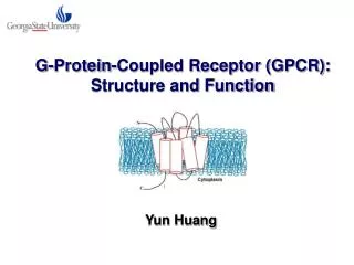
G-Protein-Coupled Receptor (GPCR): Structure and Function. Yun Huang. G-Protein-Coupled Receptor (GPCR). The largest family of integral membrane protein involved in many biological process and pathologies;

E N D
G-Protein-Coupled Receptor (GPCR): Structure and Function Yun Huang
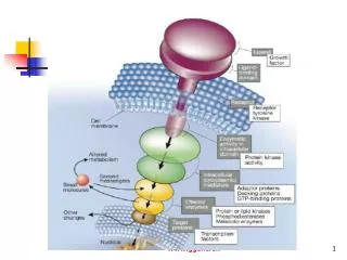
G-Protein-Coupled Receptor (GPCR) • The largest family of integral membrane protein involved in many biological process and pathologies; • 50% of all modern drugs and 25% of the top 200 best selling drugs are estimated to target GPCRs; • Transduce the signals mediated by diverse signaling molecules, such asions, peptides, lipids and photons, to induce different intracelluar function; • Bind their ligand and to activate different G proteins; http://www.sigmaaldrich.com/Area_of_Interest/Life_Science/Cell_Signaling/Scientific_Resources/Pathway_Slides___Charts.html
GPCR-Gα Fusion Protein • Upper: Schematic of a G-protein-coupled receptor (GPCR)-Gα fusion protein; • Left: G-protein cycling. Rate-limiting receptor-promoted GDP dissociation; Nat Rev Drug Discov. 2004 Jul;3(7):575, 577-626. Review.
Three Families of GPCR • Ligands: neurotranmitters (dopamine and serotonin) • Ligands: hormones (glucagon, secretin, PTH) • Example: mGluR, CaR Nat Rev Drug Discov. 2004 Jul;3(7):575, 577-626. Review.
Family 1 GPCRs: Rhodopsin-like Receptors • Represent the predominant class of GPCRs; • Several highly conserved amino acid (red circles); • Disulfide bridge between first and second extracellular loops (ECLs), palmitoylated cys in C-tail; • The binding of small molecule ligands occures within the TM region; • Example: Rhodopsin, Dopamine receptor, Chemokine receptors; Nat Rev Drug Discov. 2004 Jul;3(7):575, 577-626. Review.
Rhodopsin N • Rod cell specific visual pigment protein found in the vertebrate retina • Responsible for achromatic vision under dim light conditions • 348 amino acids, 7-TM, 11-cis-retinaldehyde (chromophore, derived from Vit A) • Posttransliational modifications: acetylation (M1), N-Glycosylation (N2, N15), S-palmitoylation (C322, C323) • Prosthetic group retinaldehyde moiety covalently attached to K296 • Functions as oligomers (dimers?) 3 4 1 5 6 2 7 C 1U19 Current Opinion inStructural Biology, 2005, 15:408-415; Experimental Eye Research, 2005, 81: 366-367
Visual Signal Transduction http://www.biocarta.com/pathfiles/m_gpcrPathway.asp
Family 2 GPCRs: Secretin-like Receptors • 15 peptide-binding receptors in human; • Relatively long N-terminus (~100-160 residues) and a juxtamembrane doamin of 7 membrane-spanning α-helices (J-domain); • Tertiary structure is stabilized by 3 disulfide bonds within 6 highly conserved Cys • Share little sequence homology with family 1 and 3 GPCRs; • Example: Corticotropin-releasing factor receptor, Glucagon receptor, PTH receptor; Nat Rev Drug Discov. 2004 Jul;3(7):575, 577-626. Review.
Family 2 GPCRs: Secretin-like Receptors Drug Discovery today, 2005: March;10(3):417-427. Review.
Corticotropin-Releasing Factor Receptor (CRF) • Related to human stress response; • NMR structure of ECD1-CRF-R2β; • Clustered in the cleft region between the tip of the first β-sheet and the edge of the “palm” of the second β-sheet; • The central core contains a salt bridge sandwiched between aromatic side chains; PNAS, 2004;101: 12836-12841.
Binding Model of Family 2 GPCRs Peptide interaction (two-domain model) Non-peptide interaction Drug Discovery today, 2005: March;10(3):417-427. Review.
Drug Design Based on Protein Structure • Comprise a central core: a “top” side chain and a “bottom” aromatic ring; • Developed by Janssen Pharmaceuticals NY & Neurocrine Biosciences; • Phase II clinical trials for treating depression and anxiety Drug Discovery today, 2005: March;10(3):417-427. Review.
Family 3 GPCRs: mGluR-like Receptors • Very long N-terminus; • Ligand-binding domain is located in N-terminus and is thought to resemble a Venus fly trap (VFT) that can open and close with the agonist bound inside; • Cys residues are conserved, the tertiary structures are likely highly conserved; • Share similar ligands and downstream signaling pathway; • Constitutive homo- or heterodimers: mGluR (homo), GAGAβ(hetero); • Example: metabotropic glutamate receptor (mGluR), CaR, GABAβ Nat Rev Drug Discov. 2004 Jul;3(7):575, 577-626. Review.
Structure of mGluR and CaR • Glutamate is a neurotransmitter in CNS and functions in long-term potentiation, learning and memory; • Metabotropic glutamate receptor (mGluR) mediate excitatory transmission on the cellular surface through initial binding of glutamate • Calcium sensing receptor (CaR) shares 27% sequence identity with (mGluR); • Response to [Ca2+]o, L-amino acids, polyamines, ionic strength and pH; • Parathyroid CaR plays a central role in systemic PTH level and Ca2+ homeostasis; Nature 2006, 407: 971-977; Current Opinion in Neurobiology 2003, 13: 271-278
Structure of mGluR and CaR mGluR ? CaR Nature 2006, 407: 971-977; Current Opinion in Neurobiology 2003, 13: 271-278
Extracellular Calcium Sensing Receptor (CaR) CaR related signaling pathway PTH: key endocrine factor in systemic Ca2+ homeostasis and involve moving Ca2+ between bone and blood; Stimulate net bone loss or formation; Cell calcium. 2004 35: 217-228. Review;
Disease Related Natural-Occurring Mutations on CaR • Familial hypocalciuric hypercalcemia (FHH), • Neonatal severe hyperparathyroidism (NSHPT), • Autosomal dominant hypoparathyroidism (ADH) http://www.casrdb.mcgill.ca/?Topic=CasrMutation
Drug Design Based on Protein Structure • Calcimimetics: • Mimic or potentiate the action of extracellular Ca2+ at the Ca2+ receptor; • Agonists or allosteric activators of the receptor; • Calcilytics: • Ca2+ receptor antagonists; Selective and does not affect the activity of mGluRs; Increase [Ca2+]i and inhibits PTH secretion; Treatment of HPTH; Drug Discovery today, 2005: March;10(3):417-427. Review.
Big questions remain for GPCRs at the structure/function interface • Determine the structures corresponding to the various states of GPCRs, especially in the active state; • Understand how GPCRs interact with and activate heterotrimeric G proteins; • Gain a molecular understanding of the mechanism of action of allosteric modulators, therefore, leads for drug development; Nat Rev Drug Discov. 2004 Jul;3(7):575, 577-626. Review.
Reference • The state of GPCR research in 2004. Nat Rev Drug Discov. 2004 Jul;3(7):575, 577-626. • Structure of rhodopsin and the metarhodopsin I photointermediate.Curr Opin Struct Biol. 2005 Aug;15(4):408-15. • Mechanisms of peptide and nonpeptide ligand binding to Class B G-protein-coupled receptors. Drug Discov Today. 2005 Mar 15;10(6):417-27. • NMR structure and peptide hormone binding site of the first extracellular domain of a type B1 G protein-coupled receptor. Proc Natl Acad Sci U S A. 2004 Aug 31;101(35):12836-41. • Structural basis of glutamate recognition by a dimeric metabotropic glutamate receptor. Nature. 2000 Oct 26;407(6807):971-7. • Extracellular Ca2+-sensing receptors--an overview. Cell Calcium. 2004 Mar;35(3):183-96.