Radiology and Diagnostic Imaging
540 likes | 1.18k Vues
Radiology and Diagnostic Imaging. CHAPTER 20. Radiology and Diagnostic Imaging Overview. X-rays High-energy electromagnetic waves Travel in straight lines Shorter wave length than visible light Able to penetrate solid materials of varying densities

Radiology and Diagnostic Imaging
E N D
Presentation Transcript
Radiology and Diagnostic Imaging CHAPTER 20
Radiology and Diagnostic Imaging Overview • X-rays • High-energy electromagnetic waves • Travel in straight lines • Shorter wave length than visible light • Able to penetrate solid materials of varying densities • Capable of exposing a photographic plate (X-ray film) • Much the same way as a camera exposes film
Radiology and Diagnostic Imaging Overview • X-rays • Used to visualize internal organs and structures of body • Provide valuable means for verifying presence of illness or disease • Radiology • Study of the diagnostic and therapeutic uses of X-rays
PROCEDURES AND TECHNIQUES Radiology and Diagnostic Imaging
Angiocardiography(Cardiac Catheterization) • Pronounced • (an-jee-oh-kar-dee-OG-rah-fee) • (CAR-dee-ak kath-eh-ter-ih-ZAY-shun) • Defined • Specialized diagnostic procedure in which a catheter is introduced into a large vein or artery • Usually of an arm or a leg, and is then threaded through circulatory system to the heart
Angiography • Pronounced • (an-jee-OG-rah-fee) • Defined • Series of X-ray films allowing visualization of internal structures after the introduction of a radiopaque substance
Cerebral Angiography • Pronounced • (seh-REE-bral an-jee-OG-rah-fee) • (SER-eh-bral an-jee-OG-rah-fee) • Defined • Injection of a radiopaque contrast medium into an arterial blood vessel (carotid, femoral, or brachial) to make visualization of the cerebral vascular system via X-ray possible
Renal Angiography • Pronounced • (REE-nal an-jee-OG-rah-fee) • Defined • X-ray visualization of internal anatomy of the renal blood vessels (blood vessels of the kidney) after injection of a contrast medium
Arteriography • Pronounced • (ar-tee-ree-OG-rah-fee) • Defined • X-ray visualization of arteries following the introduction of a radiopaque contrast medium into the bloodstream through a specific vessel by way of a catheter
Arthrography • Pronounced • (ar-THROG-rah-fee) • Defined • Process of taking X-rays of the inside of a joint, after a contrast medium has been injected into the joint • Contrast medium makes the inside of the joint visible
Barium Enema (BE) • Pronounced • (BAH-ree-um EN-eh-mah) • Defined • Infusion of a radiopaque contrast medium, barium sulfate, into the rectum • Contrast medium is retained in lower intestinal tract while X-ray films are obtained of the lower GI tract
Barium Swallow(Upper GI Series) • Pronounced • (BAH-ree-um SWALL-oh) • Defined • Oral administration of a radiopaque contrast medium, barium sulfate, which flows into the esophagus as the person swallows • X-rays are taken as barium sulfate flows into the upper GI tract
Bronchography • Pronounced • (brong-KOG-rah-fee) • Defined • Bronchial examination via X-ray following the coating of the bronchi with a radiopaque substance
Cholangiography(Intravenous) • Pronounced • (koh-lan-jee-OG-rah-fee) • (in-trah-VEE-nus) • Defined • Visualizing and outlining of the major bile ducts following an intravenous injection of a contrast medium
Cholangiography(Percutaneous Transhepatic) • Pronounced • (koh-lan-jee-OG-rah-fee) • (per-kyoo-TAY-nee-us trans-heh-PAT-ik) • Defined • Examination of bile duct structure using a needle to pass directly into an intrahepatic bile duct to inject a contrast medium • Also known as PTC or PTHC
Cholangiopancreatography(Endoscopic Retrograde) • Pronounced • (koh-lan-jee-oh-pan-kree-ah-TOG-rah-fee) • (en-doh-SKOP-ic RET-roh-grayd) • Defined • Procedure that examines the size of and the filling of the pancreatic and biliary ducts through direct radiographic visualization with a fiberoptic endoscope
Cholecystography(Oral) • Pronounced • (koh-lee-sis-TOG-rah-fee) • Defined • Visualization of the gallbladder through X-ray following the oral ingestion of pills containing a radiopaque iodinated dye
Cineradiography • Pronounced • (sin-eh-ray-dee-OG-rah-fee) • Defined • Diagnostic technique combining the techniques of fluoroscopy, radiography, and cinematography by filming the images that develop on a fluorescent screen with a movie camera
Computed Axial Tomography (CT, CAT) • Pronounced • (kom-PEW-ted AK-see-al toh-MOG-rah-fee) • Defined • Painless, noninvasive diagnostic X-ray procedure using ionizing radiation that produces a cross-sectional image of the body
Voiding Cystourethrography • Pronounced • (VOYD-ing sis-toh-yoo-ree-THROG-rah-fee) • Defined • X-ray visualization of the bladder and urethra during the voiding process, after the bladder has been filled with a contrast material
Digital Subtraction Angiography (DSA) • Pronounced • (DIJ-ih-tal sub-TRAK-shun an-jee-OG-rah-fee) • Defined • X-ray images of blood vessels only, appearing without any background, due to the use of a computerized digital video subtraction process
Echocardiography • Pronounced • (ek-oh-kar-dee-OG-rah-fee) • Defined • Diagnostic procedure for studying the structure and motion of the heart via ultrasound • Useful in evaluating structural and functional changes in a variety of heart disorders
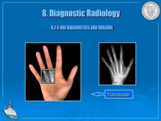
Fluoroscopy • Pronounced • (floor-or-OSS-koh-pee) • Defined • Radiological technique used to examine the function of an organ or a body part using a fluoroscope
Hysterosalpingography • Pronounced • (his-ter-oh-sal-ping-OG-rah-fee) • Defined • X-ray assessment of uterus and fallopian tubes by injecting a contrast material into these structures
Lymphangiography • Pronounced • (lim-fan-jee-OG-rah-fee) • Defined • X-ray assessment of lymphatic system following injection of a contrast medium into lymph vessels in the hand or foot
Magnetic Resonance Imaging (MRI) • Pronounced • (mag-NET-ik REZ-oh-nans IM-ij-ing) • Defined • Noninvasive scanning procedure that provides visualization of fluid, soft tissue, and bony structures without the use of radiation
Mammography • Pronounced • (mam-OG-rah-fee) • Defined • Process of taking X-rays of the soft tissue of the breast to detect various benign and/or malignant growths before they can be felt
Myelography • Pronounced • (my-eh-LOG-rah-fee) • Defined • Introduction of contrast medium into the lumbar subarachnoid space through a lumbar puncture to visualize the spinal cord and vertebral canal through X-ray examination
Positron Emission Tomography Scan (PET) • Pronounced • (POZ-ih-tron ee-MISH-un toh-MOG-rah-fee) • Defined • Noninvasive diagnostic imaging method that demonstrates the biological function of the body before anatomical changes take place • Scan produces computerized radiographic images of the body structures when radioactive substances are administered to the patient • Substances are inhaled or injected
Pyelography(Intravenous) (IVP) • Pronounced • (pye-eh-LOG-rah-fee) • (in-trah-VEE-nus) • Defined • Radiographic procedure that provides visualization of the entire urinary tract: kidneys, ureters, bladder, and urethra • Also known as intravenous pyelogram or excretory urogram
Radiation Therapy • Pronounced • (ray-dee-AY-shun THAIR-ah-pee) • Defined • Delivery of ionizing radiation to accomplish one or more of the following: • Destruction of tumor cells • Reduction of tumor size • Decrease in pain • Relief of obstruction • To slow or stop the spread of cancer cells
Radiation Therapy • Radiation therapy • Destroys rapidly multiplying cells regardless of whether they are cancerous • Goal is to reach maximum tumor control with no, or minimal, normal tissue damage • May be delivered by teletherapy (external) • May be delivered by brachytherapy (internal)
Radioactive Iodine Uptake • Pronounced • (ray-dee-oh-AK-tiv EYE-oh-dine UP-tayk) • Defined • Examination that determines the position, size, shape, and physiological function of the thyroid gland through the use of radionuclear scanning • Image of the thyroid is recorded and visualized after a radioactive substance is given
Scanning (Bone, Brain, Liver, Lungs) • Pronounced • (SCAN-ing) • Defined • Scanning is the process of recording emission of radioactive waves, using a gamma camera (scanner) • After an intravenous injection of a radionuclide material into the particular part of the body being studied
Scanning (Bone, Brain, Liver, Lungs) • Defined • Image of the area being studied is displayed by recording concentration or collection of a radioactive substance specifically drawn to that area
Scanning • Bone • Involves intravenous injection of a radionuclide material absorbed by bone tissue • Used to detect spread of cancer to the bones, osteomyelitis, and other destructive changes in the bones
Scanning • Brain • Nuclear scanning of cranial contents two hours after an intravenous injection of radioisotopes • Useful in diagnosing abnormal findings such as an acute cerebral infarction, cerebral neoplasm, cerebral hemorrhage, brain abscess, aneurysms, cerebral thrombosis, hematomas, hydrocephalus, cancer metastasis to the brain, and bleeds
Scanning • Liver • Noninvasive scanning technique that enables the visualization of the shape, size, and consistency of the liver after the IV injection of a radioactive compound • Useful in detecting cysts, abscesses, tumors, granulomas, or diffuse infiltrative processes affecting the liver
Scanning • Lung • Visual imaging of the distribution of ventilation or blood flow in the lungs by scanning the lungs after the patient has been injected with or has inhaled radioactive material
Scanning • Spleen • Noninvasive scanning technique that enables the visualization of the shape, size, and consistency of the spleen after injection of radioactive red blood cells • Useful in detecting damage, tumors, and other problems
Single-Photon Emission Computed Tomography (SPECT) • Pronounced • (single FOH-ton ee-MISH-un kom-PEW-ted toh-MOG-rah-fee) • Defined • Nuclear imaging procedure that shows how blood flows to tissues and organs • Tracking of radioactive material allows physician to see perfusion of blood to tissues and organs
Small Bowel Follow-Through • Pronounced • (Small Bowel Follow-Through) • Defined • Oral administration of a radiopaque contrast medium, barium sulfate, which flows through the GI system • X-ray films are obtained at timed intervals to observe progression of barium through small intestines
Tomography • Pronounced • (toh-MOG-rah-fee) • Defined • X-ray technique used to construct a detailed cross-section, at a predetermined depth, of a tissue structure • Useful in identifying space-occupying lesions in the liver, brain, pancreas, and gallbladder
Ultrasonography(Ultrasound) • Pronounced • (ull-trah-son-OG-rah-fee) • Defined • Procedure in which sound waves are transmitted into body structures as a small transducer is passed over the patient’s skin • Sound waves are reflected back into the transducer and are interpreted by a computer that converts waves to a composite picture form
Ultrasonography • Abdominal ultrasound • Use of reflected sound waves to provide reliable visualization of the liver, gallbladder, bile ducts, pancreas, kidneys, bladder, and ureters
Ultrasonography • Pelvic ultrasound • Noninvasive procedure that uses high-frequency sound waves to examine the abdomen and pelvis • Can be used to locate a pelvic mass, an ectopic pregnancy, or an intrauterine device, and to inspect and assess the uterus, ovaries, and fallopian tubes
Ultrasonography • Renal ultrasound • Noninvasive ultrasound of the kidneys that is useful in distinguishing between fluid-filled cysts and solid masses, detecting renal calculi, identifying obstructions, and evaluating transplanted kidneys • Thyroid Echogram (ultrasound) • Ultrasound examination important in distinguishing solid thyroid nodules from cystic nodules
Venography • Pronounced • (vee-NOG-rah-fee) • Defined • Technique used to prepare an X-ray image of veins • Veins are injected with a radiopaque contrast medium • Phlebography
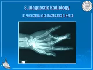
X-rays • Pronounced • (ECKS-rays) • Defined • Use of high-energy electromagnetic waves, passing through the body onto a photographic film, to produce a picture of the internal structures of the body for diagnosis and therapy