
Image Analysis and Gene Expression Data Preprocessing
E N D
Presentation Transcript
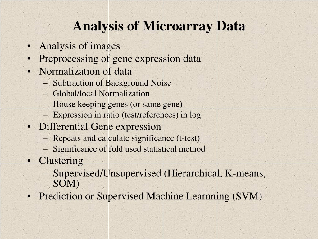
Analysis of Microarray Data • Analysis of images • Preprocessing of gene expression data • Normalization of data • Subtraction of Background Noise • Global/local Normalization • House keeping genes (or same gene) • Expression in ratio (test/references) in log • Differential Gene expression • Repeats and calculate significance (t-test) • Significance of fold used statistical method • Clustering • Supervised/Unsupervised (Hierarchical, K-means, SOM) • Prediction or Supervised Machine Learnning (SVM)
pseudo-colourimage sample(labelled) probe (on chip) Technical
Images from scanner • Resolution • standard 10m [currently, max 5m] • 100m spot on chip = 10 pixels in diameter • Image format • TIFF (tagged image file format) 16 bit (65’536 levels of grey) • 1cm x 1cm image at 16 bit = 2Mb (uncompressed) • other formats exist e.g.. SCN (used at Stanford University) • Separate image for each fluorescent sample • channel 1, channel 2, etc.
Images in analysis software • The two 16-bit images (Cy3, Cy5) are compressed into 8-bit images • Display fluorescence intensities for both wavelengths using a 24-bit RGB overlay image • RGB image : • Blue values (B) are set to 0 • Red values (R) are used for Cy5 intensities • Green values (G) are used for Cy3 intensities • Qualitative representation of results
Pseudo-colour overlay Cy3 Cy5 Images : examples
Processing of images • Addressing or gridding • Assigning coordinates to each of the spots • Segmentation • Classification of pixels either as foreground or as background • Intensity determination for each spot • Foreground fluorescence intensity pairs (R, G) • Background intensities • Quality measures
Background intensity • Spot’s measured intensity includes a contribution of non-specific hybridization and other chemicals on the glass • Fluorescence from regions not occupied by DNA should by different from regions occupied by DNA -> one solution is to use local negative controls (spotted DNA that should not hybridize) • Different background methods : • Local background • Morphological opening • Constant background • No adjustment
ScanAlyze ImaGene Spot, GenePix Local background • Focusing on small regions surrounding the spot mask. • Median of pixel values in this region • Most software package implement such an approach • By not considering the pixels immediately surrounding the spots, the background estimate is less sensitive to the performance of the segmentation procedure
Morphological opening • Non-linear filtering, used in Spot • Use a square structuring element with side length at least twice as large as the spot separation distance • Compute local minimum filter, then compute local maximum filter • This removes all the spots and generates an image that is an estimate of the background for the entire slide • For individual spots, the background is estimated by sampling this background image at the nominal center of the spot • Lower background estimate and less variable
Constant background • Global method which subtracts a constant background for all spots • Some evidence that the binding of fluorescent dyes to ‘negative control spots’ is lower than the binding to the glass slide • -> More meaningful to estimate background based on a set of negative control spots • If no negative control spots :approximation of the average background =third percentile of all the spot foreground values
No background adjustment • Do not consider the background • Probably not accurate, but may be better than some forms of local background determination!
Histograms Signal/Noise = log2(spot intensity/background intensity)
Preprocessing of Gene expression Data • Scale transformation • CY3/CY5 • LOG(CY3/CY5) • Replicates handling • Inconsistent replicate removal • Replicate merging • Missing value handling • Removal of patterns having excess of missing values • Value of missing points • Flat pattern filtering • Unknown Gene Removing
Preprocessing: Normalization • Why? To correct for systematic differences between samples on the same slide, or between slides, which do not represent true biological variation between samples. • How do we know it is necessary? By examining self-self hybridizations, where no true differential expression is occurring. We find dye biases which vary with overall spot intensity, location on the array, plate origin, pins, scanning parameters,….
Normalization Techniques • Global normalization • Divide channel value by means • Control spots • Common spots in both channels • House keeping genes • Ratio of intensity of same gene in two channel is used for correction • Iterative linear regression • Parametric nonlinear nomalization • log(CY3/CY5) vs log(CY5)) • Fitted log ratio – observed log ratio • General Non Linear Normalization • LOESS • curve between log(R/G) vs log(sqrt(R.G))
Pre-processed cDNA Gene Expression Data Slides On p genes for n slides: p is O(10,000), n is O(10-100), but growing, slide 1 slide 2 slide 3 slide 4 slide 5 … 1 0.46 0.30 0.80 1.51 0.90 ... 2 -0.10 0.49 0.24 0.06 0.46 ... 3 0.15 0.74 0.04 0.10 0.20 ... 4 -0.45 -1.03 -0.79 -0.56 -0.32 ... 5 -0.06 1.06 1.35 1.09 -1.09 ... Genes Gene expression level of gene 5 in slide 4 = Log2(Red intensity / Green intensity) These values are conventionally displayed on a red(>0)yellow (0)green (<0) scale.
Scatterplots: always log, always rotate log2R vs log2G M=log2R/G vs A=log2√RG
Classification • Task: assign objects to classes (groups) on the basis of measurements made on the objects • Unsupervised: classes unknown, want to discover them from the data (cluster analysis) • Supervised: classes are predefined, want to use a (training or learning) set of labeled objects to form a classifier for classification of future observations
Cluster analysis • Used to find groups of objects when not already known • “Unsupervised learning” • Associated with each object is a set of measurements (the feature vector) • Aim is to identify groups of similar objects on the basis of the observed measurements
Example: Tumor Classification • Reliable and precise classification essential for successful cancer treatment • Current methods for classifying human malignancies rely on a variety of morphological, clinical and molecular variables • Uncertainties in diagnosis remain; likely that existing classes are heterogeneous • Characterize molecular variations among tumors by monitoring gene expression (microarray) • Hope: that microarrays will lead to more reliable tumor classification (and therefore more appropriate treatments and better outcomes)
Nearest Neighbor Classification • Based on a measure of distance between observations (e.g. Euclidean distance or one minus correlation) • k-nearest neighbor rule (Fix and Hodges (1951)) classifies an observation X as follows: • find thek observations in the learning set closest to X • predict the class of X by majority vote, i.e., choose the class that is most common among those k observations. • The number of neighbors kcan be chosen by cross-validation
Hierarchical Clustering • Produce a dendrogram • Avoid prespecification of the number of clusters K • The tree can be built in two distinct ways: • Bottom-up: agglomerative clustering • Top-down: divisive clustering
Partitioning vs. Hierarchical • Partitioning • Advantage: Provides clusters that satisfy some optimality criterion (approximately) • Disadvantages: Need initial K, long computation time • Hierarchical • Advantage: Fast computation (agglomerative) • Disadvantages: Rigid, cannot correct later for erroneous decisions made earlier
Issues in Clustering • Pre-processing (Image analysis and Normalization) • Which genes (variables) are used • Which samples are used • Which distance measure is used • Which algorithm is applied • How to decide the number of clusters K
Filtering Genes • All genes (i.e. don’t filter any) • At least k (or a proportion p) of the samples must have expression values larger than some specified amount, A • Genes showing “sufficient” variation • a gap of size A in the central portion of the data • a interquartile range of at least B • Filter based on statistical comparison • t-test • ANOVA • Cox model, etc.
Average linkage hierarchical clustering, melanoma only unclustered ‘cluster’