Download
1 / 9
90 likes | 109 Vues
Astroglial cradle in the life of the synapse

E N D
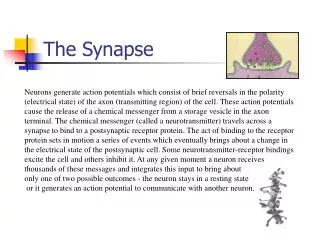
Astroglial cradle in the life of the synapse Alexei Verkhratsky1,2,3and Maiken Nedergaard4 1Faculty of Life Sciences, University of Manchester, Manchester, UK 2Achucarro Center for Neuroscience, IKERBASQUE, Basque Foundation for Science, 48011 Bilbao, Spain 3University of Nizhny Novgorod, Nizhny Novgorod 603022, Russia 4Division of Glia Disease and Therapeutics, Center for Translational Neuromedicine, University of Rochester Medical School, Rochester, NY 14580, USA rstb.royalsocietypublishing.org Astroglial perisynaptic sheath covers the majority of synapses in the central nervous system. This glial coverage evolved as a part of the synaptic structure in which elements directly responsible for neurotransmission (exocytotic machinery and appropriate receptors) concentrate in neuronal membranes, whereas multiple molecules imperative for homeostatic maintenance of the synapse(transporters forneurotransmitters,ions,aminoacids, etc.)are shifted to glial membranes that have substantially larger surface area. The astrocytic perisynapticprocesses actasan‘astroglial cradle’ essentialforsynaptogenesis, maturation, isolation and maintenance of synapses, representing the funda- mental mechanism contributing to synaptic connectivity, synaptic plasticity and information processing in the nervous system. Review Cite this article: Verkhratsky A, Nedergaard M. 2014 Astroglial cradle in the life of the synapse. Phil. Trans. R. Soc. B 369: 20130595. http://dx.doi.org/10.1098/rstb.2013.0595 One contribution of 23 to a Theme Issue ‘Brain circuitry outside the synaptic cleft’. 1. Multiple components of the central synapse Thepowerofthe brain lies inthe intercellularconnections; some tens oftrillions of theseconnectionscreatetheneuralwebthatisthesubstrateforinformationproces- sing. The connectivity of neural networks is immensely plastic and it is constantly remodelled under the influence of the environment and experience; this remark- able plasticity underlies learning. Intercellular connections in the nervous system are represented by chemical and electrical synapses, with the former predomi- nantly established between neurons and the latter between neuroglia. Chemical transmission in the brain is not confined to synapses, it also operates in a ‘volume transmission’ mode when neurotransmitters diffuse through interstitial space, finding their multiple targets between neurons and neuroglial cells. Synaptic structures evolved over the last approximately 600 million years, after the first diffuse nervous system appeared in primitive animals such as hydras and comb jellies; this first nervous system was composed from cells of a single cell type, the neurons, which establish a neuronal net through synaptic contacts. Subsequent evolution of the nervous system progressed by the way of great diversification and specialization of cells. The first glial-like cells emerged in nematodes, they became specialized in annelids and attained a high degree of morphological and functional heterogeneity in arthropods. In basal chor- dates, the new type of glial cells, the radial glia, replaced parenchymal glial cells, which signalled an advent of layered organization of the central nervous system (CNS). Increase in the size of the CNS instigated re-emergence and diver- sificationofastrocytesandappearanceofmyelin[1].Thebrainandthespinalcord of mammals contain hundreds of distinct types of neurons and many types of neuroglia. Similarly, there are many types of synapses established between neur- onalterminalsandeffectororgansintheperipheryorbetweenneuronalterminals and other neurons and between neurons and some NG2 glial cells [2] in the CNS as well as in sympathetic ganglia or enteric plexuses. Synapses in the CNS are composed of several distinct components (figure 1), which include (i) the presynaptic terminal, (ii) the postsynaptic element that can be represented, for example, by the dendritic spine, (iii) the perisynaptic process of the astrocyte, (iv) theprocessofa neighbouringmicroglialcellthat periodically contacts the synaptic structure and (v) the extracellular matrix (ECM), which is present in the synaptic cleft and also extends extra-synaptically. The concept of this multi-partite synaptic assembly evolved over a recent decade, when it became clear that complex multidirectional relations exist between all the Subject Areas: neuroscience Keywords: astroglia, synapse, astroglial cradle, synaptogenesis, neurotransmission, potassium buffering Authors for correspondence: Alexei Verkhratsky e-mail: alexej.verkhratsky@manchester.ac.uk Maiken Nedergaard e-mail: nedergaard@urmc.rochester.edu & 2014 The Author(s) Published by the Royal Society. All rights reserved.
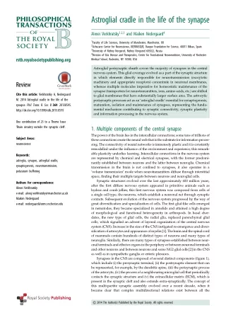
presynaptic terminal 2 rstb.royalsocietypublishing.org synaptogenesis synaptic isolation synaptic maturation synaptic maintenance neurotransmitter homeostasis ion homeostasis ECM volume homeostasis Phil. Trans. R. Soc. B 369: 20130595 astroglial perisynaptic sheath synaptic extinction (?) microglial process postsynaptic neuron Figure 1. Astroglial cradle embraces and fosters multi-partite synapse in the CNS. The majority of synapses in the brain and spinal cord are composed of several components that include: the presynaptic terminal; the postsynaptic part; the perisynaptic process of the astrocyte; the process of a neighbouring microglial cell that periodically contacts the synaptic structure; and the ECM present in the synaptic cleft and also extending extra-synaptically. Astroglial perisynaptic sheaths enwrap synaptic structures and regulate, influence and assist synaptogenesis, synaptic maturation, synaptic maintenance and synaptic extinction. (Online version in colour.) components outlined above [3–8]. In physiology, the concept of the multi-partite synapse (mainly in its ‘tripartite’ version) is linked to the idea of fast ‘gliotransmission’, which postulates that Ca2þ-regulated exocytotic release of neurotransmitters from astrocytes directly contributes to synaptic transmission. The gliotransmission has recently been overviewed in detail [9], although its importance for synaptic physiology remains a controversy [7,10–13]. Debates on this matter, which have generated much emotion in the neuroglial field, are outside the scope of the current narrative, although we are favouring the notion that fast gliotransmission plays little role in physiological activity of multi-partite synapse. Each and everyone of the synaptic structural components plays a distinct role in the life of the synapse, from its appear- ance and maturation to its maintenance and, if needed, to its extinction. The microglia cells appear in the CNS from the very early embryonic period [14], and, being essentially the firstglialcellsexistinginthedevelopingCNS,possiblycontrib- ute to early synaptogenesis; at the later developmental stages, microglial cells are fundamental for shaping synaptic connec- tivity through, for example, synaptic pruning/stripping [15,16]. Similarly, the ECM regulates synaptogenesis, intercon- nects pre- and postsynaptic protein complexes, controls receptor trafficking through integrins-mediated signalling and modulates postsynaptic excitability [5,17–20]. In this brief essay, we shall overview the contribution of astroglia to the life of a synapse in the CNS. processes (known as PAPs); these latter are identified by immu- noreactivitytoglutamatesynthetaseandglutamatetransporters andaredevoidofglialfibrillaryacidicprotein(GFAP)[21,22].In total,about60%ofsynapsesintheCA1areaofthehippocampus are enwrapped with membranes of protoplasmic astrocytes; furthermore, this coverage seems to vary between different synapses: astroglial processes surround approximately 90% of all large mushroom spines and perforated synapses [23], whereas only 50% of small macular synapses are enclosed with astroglia. In the cerebellum, almost all of the synapses formed by parallel fibres on the dendrites of Purkinje neuron are tightly covered by terminal processes of Bergmann glial cells; these glial structures are quite complex, sprouting from specialized appendages that protrude from the main shaft of Bergmann glial processes [24]. The perisynaptic astroglial sheaths covering synapses are exceedingly thin, the profiles of these perisynaptic structures being on average less than 200 nm (and often less than 100 nm) in diameter [22]. Impor- tantly, astroglial PAPs demonstrate remarkable morphological plasticity, whenthroughretractingorextendingastroglialmem- branes the degree of synaptic coverage can be dynamically regulated [25]. It has to be noted, however, that the data on synapticcoveragebyastroglialprocessesremaingenerallyinsuf- ficient with only a few brain regions being investigated; the detailed cartography of astroglial synaptic profiles throughout the CNS is very much longed for. (b) Physiology The perisynaptic astroglial membrane represents the major part of the cell surface area, accounting for approximately 80% of the total astroglial plasmalemma, and yet it contributes only a minor fraction (approx. 4–10%) to the cellular volume, this being reflected by an extremely high surface-to-volume ratio (around 2521mm [26]). This in essence means that the 2. Astroglial synaptic coverage (a) Morphology ThemajorityofsynapsesintheCNSarecoveredwithperisynap- tic astroglial sheaths that emanate from peripheral astroglial
potassium homeostasis 3 rstb.royalsocietypublishing.org Kir4.1 NKA KCC1 K+ 2K+ 1K+ 1Cl– 3Na+ K+ Phil. Trans. R. Soc. B 369: 20130595 neurotransmitter homeostasis Xc– EAAT1,2 GAT1,3 GlyT1 SN1,2 ENT1 1Cl– 1Cl– 1Glu– 3Na+ 1H+ 1CYS GABA 2Na+ GLY 2Na+ 1H+ ADO 1GLN1GLN 1K+ 1Na+ pH calcium homeostasis metabolic support ROS homeostasis homeostasis NHE NBC NCX MCT1 NAAT 1Ca2+ 2/3HCO3– 1Na+ 3Na+ 2Na+ 1Na+ 1ASC lactate 1H+ 1Ca2+ 3Na+ direct mode reverse mode Figure 2. Homeostatic molecular cascades localized in astroglial perisynaptic processes. (Online version in colour.) perisynaptic astroglial plasmalemma with very little cytosol. Such a morphological arrangement provides an excessive space for receptors, chan- nels, transporters and pumps that contribute to dynamic neuronal–glial signalling and accomplish glial homeostatic function;atthesametime,shiftingofallhomeostaticmolecules to glial membranes allows maximal density of exocytotic machinery in the presynapse and neurotransmitter receptors in the postsynaptic membrane. Astrocytes possess a highly heterogeneous complement of neurotransmitter receptors (both iono- and metabotropic), expression of which varies between brain regions, being most probably regulated by the local chemical environment [27]. Astroglial ionotropic recep- tors are activated by neurotransmitters released in the course of synaptic transmission and create local transient micro- domains of high concentration of cytosolic Ca2þand Naþ that represent a substrate for glial excitability. The perisynaptic processes also contain numerous transporters critical for homeostatic transport of ions, neurotransmitters, glutamine, lactate, etc. (figure 2). These transporters create substantial ion fluxes; the PAPs and perisynaptic astroglial compartments membrane sheath is limited to are generally poor in organelles [22], and in particular they are almost completely devoid of endoplasmic reticulum [28] making transmembrane ion fluxes fundamental for local ion signalling mediated by both Ca2þand Naþ[29,30]. 3. Astrocytes cradle: fostering and maintaining synaptic connectivity The intimate relationship between perisynaptic astroglial com- partments and central synapses is synthesized in a concept of the astroglial cradle [7], which appears as a fundamental elementinthelifeofasynapse,beinginvolvedinsynapticgen- esis and maturation and providing lifelong support for the synaptic function (figure 1). (a) Synaptogenesis and synaptic maturation The life of the synapse progresses through several stages that include (i) formation of an initial contact between the term- inal and postsynaptic neuron, (ii) maturation of the synapse, (iii) its stabilization and maintenance and (iv) elimination.
[40]. The astroglial Kþremoval system is represented by seve- ral complementary molecular Naþ/KþATPase, inward rectifying Kir4.1 channels, Kþ/Cl2 co-transporter KCC1 (SLC12A4) transporterNKCC1(SLC12A2)[41,42].Morerecentdatasuggest that inhibition of Naþ/KþATPase reduced post-stimulus clear- ance of Kþtransients, whereas inhibition of Kir4.1 channels and NKCC1 had minor effects [43]. The transporters are regulated by [Naþ]iand (indirectly) by [Ca2þ]i: for example, increase in astroglial Ca2þactivates the forward mode of the Naþ/Ca2þ exchanger that mediates substantial Naþinflux, which in turn stimulates Naþ/KþATPase and alters the Naþelectro-driving forcethatmaintainsfunctionalactivityofrelevantSLCs[44].Pot- assium buffering not only removes excess Kþbut also affects short-term plasticity for example in hippocampal synapses [44,45]. Potassium ions, accumulated by astrocytes, can sub- sequently dissipate through astroglial syncytium through the mechanism known as spatial Kþbuffering [46]. Once formed, synapses survive for several hours in the early postnatal period and for days and month in the adult CNS [31]. The major wave of genesis of glutamatergic synapses in themammalianbrainoccurs intheearlypostnatalperiod (post- natal weeks 1–3 in rodents); this wave of synaptogenesis immediately follows massive astrogliogenesis [32]. Astroglia contribute to synaptogenesis through secreting various factors, in particular cholesterol [33] and thrombospondins; the latter are an astroglia-derived component of ECM. Thrombospondins exert their effects through an accessory subunit of voltage-acti- vated calcium channels identified as a2/d1 protein (or CaCna2d1) [18]. Astrocytes may also promote synaptogenesis through oestradiol, g-protocadherins, integrins and protein kinase C [34]. Not all central synapses require glia, and for instance inhibitory GABA-ergic contacts develop before the profuse appearance of astrocytes [35]. Similarly, factors produced and secreted by astrocytes enhance and facilitate maturation of synapses affecting both pre- and postsynaptic neuronal compartments. These astroglia- derived molecules include activity-dependent neurotrophic factor and tumour necrosis factor awhich regulate the traffick- ing of glutamate receptors into postsynaptic membranes, whereas cholesterol may enhance neurotransmitter release from presynaptic terminals [36]. Astrocytes may also regulate the number of synapses on the given neuron by denying presynaptic terminals the con- tact with postsynaptic membranes and can be involved in synapse elimination. In particular, astrocytes in vitro have been found to label synaptic terminals with complement factor C1q, this tag being recognized by microglia with sub- sequent selective phagocytotic removal of the synaptic contact [36,37]. Other mechanisms may also exist, although the overall role and contribution of astrocytes to synaptic extinction remains controversial [38]. 4 mechanisms that include rstb.royalsocietypublishing.org Naþ/Kþ/Cl2 and co- Phil. Trans. R. Soc. B 369: 20130595 (ii) Proton homeostasis and pH regulation Astrocytes are indispensable for regulation of pH in the CNS interstitium by providing transmembrane transport for Hþ(by sodium–proton exchanger, SLC9A1/NHE-1) and bicarbonate (through sodium–bicarbonate co-transporter SLC4A4/NBC). In addition, astrocytes contribute to extracellular Hþlevels through the lactate transporter SLC16A1/MCT-1, which co-transports 1 Hþwith 1 lactate, and through glutamate trans- porters,whichremove1Hþfromtheextracellularspacetogether with each glutamate molecule (see [47] for details). (iii) Other ions Astrocytes contribute to homeostasis of other biologically relevant ions. In particular, activation of astroglial anion channels trigger Cl2efflux (because of high cytosolic Cl2con- centration) that is activated for example in hypo-osmotic stress. Astrocytes can also accumulate Cl2by NKCC1. Astro- cytes participate in Zn2þhomeostasis [48], being endowed with ZnTs/SLC30 transporter; incidentally the lattermay med- iate astroglial Zn2þrelease in hypo-osmotic conditions [49]. Finallyastrocytes,may,atleastinprinciple,contributetorestor- ation of extracellular Ca2þin the cleft after periods of intense neuronal activity that may substantially decrease [Ca2þ]i[50]. Lowering extracellular Ca2þcan trigger astroglial intracellular Ca2þrelease [51]; and Ca2þmay subsequently leave the astrocyte through the Naþ/Ca2þexchanger. (b) Synaptic isolation A fundamental function of the astroglial cradle is synaptic iso- lation, when perisynaptic astroglial sheath shields the synapse fromtheouterspace,preventingboth‘spill-over’ofthetransmit- ters from the synaptic cleft and ‘spill-in’ of the transmitters into thecleftfromtheextra-synapticstructures[7].Thisideaofsynap- tic isolation or insulation has a long history, being invented by Santiago Ramon y Cajal (who credited his brother Pedro with the suggestion [39]). The astroglial cradle isolates synapses both mechanically (by enwrapping synaptic structures with perisynapticmembranes)andfunctionallybyactiveneurotrans- mitter uptake,althoughofcoursenot allthesynapsesarefenced by astroglial membranes and the functional consequences for these ‘open’ synapses remain to be investigated. (d) Neurotransmitter homeostasis (i) Glutamate Astroglial cells are fundamental elements of glutamatergic transmission in the CNS that control the extracellular distri- bution of glutamate and maintain the neuronal glutamate pool by supplying glutamine. Astrocytes regulate extracellular glutamate concentration by balancing glutamate uptake through Naþ-dependent astroglia-specific glutamate transpor- ters (SLC1A3/EAAT1 and SLC1A2/ EAAT2) and glutamate release mainly through the cysteine–glutamate exchanger or systemXc2(SLC7A11)[52,53]. Thesetwo typesof transporters are differentially expressed in the astroglial membranes so that glutamate uptake dominates around the synaptic cleft, whereas cysteine–glutamate exchangers are concentrated in extra-synaptic regions. In the synaptic cleft at rest, glutamate (c) Ion homeostasis Ion homeostasis of the extracellular space is of paramount importance for CNS function; fluctuations in extracellular ion concentrations rapidly affect excitability of neurons that may cause numerous adverse effects. This is particularly important for Kþions that travel across neuronal membranes firing action potentials and postsynaptic membranes activated by neurotransmitters. (i) Potassium buffering Astrocytes are central for extracellular Kþhomeostasis in the CNS, their critical role being recognized by Leif Hertz in 1965
is kept at low nanomolar levels (that prevents desensitization of glutamate receptors) and a high density of glutamate trans- porters ascertains rapid clearance of glutamate upon synaptic transmission. Extra-synaptically, however, glutamate is main- tained at low micromolar concentrations that provide for tonic modulation of synaptic transmission and plasticity through extra-synaptic metabotropic (and possibly NMDA) glutamate receptors [53]. Besides providing for glutamate clearance and controll- ing interstitial glutamate distribution, astrocytes maintain glutamatergic transmission through supplying neurons with glutamate precursor, glutamine. The latter is synthesized solely by astroglia-specific glutamine synthetase and is fed to neurons through a coordinated amino acid transporting sys- tem. Astrocytes express Naþ/Hþ-dependent sodium-coupled neutral amino acid transporters and SN2/SNAT5/SLC38A5, which mediate glutamine efflux, whereas neurons specifically express the sodium-coupled neutral amino acid transporters ATA1/SNAT1/SLC38A1 and ATA2/SNAT2/SLC38A2, which act as influx transporters mediating glutamine accumulation into the neuronal com- partment [54]. Glutamate uptake, glutamate conversion by glutamine synthetase and glutamine transport together rep- resent the glutamate–glutamine shuttle that maintains a releasable pool of neuronal glutamate and sustains glutamater- gic transmission [55,56]. Release of glutamine by astrocytes is linked to synaptic activity; increase in synaptically released glutamate increases astroglial release of glutamine, this process possibly being mediated through activation of glutamate trans- porters and consequent increase in cytosolic Naþconcentration in perisynaptic astroglial processes [57,58]. Inhibition of this pathway affects glutamatergic trans- mission differentially in different brain regions: for example, in the retina neuronal responses disappear in 2 min after inhi- bition of glutamine synthetase, whereas in hippocampus glutamatergic activity lasts for some hours after cessation of glutamine supply [59]. transporters and is catabolized intracellularly by adenosine kinase that in the CNS is expressed exclusively in astrocytes [63]. Whether astrocytes or neurons represent the main source for extracellular adenosine (either released directly or derived from degradation of ATP) remains a matter of debate [64,65]. Nonetheless, astrocytes are most likely the main sink foradenosine because adenosine kinase activity keeps cytosolic adenosine levels low, thus allowing equilibrium transporter to generate adenosine influx. This is a vital function, and transgenic deletion of adenosine kinase is lethal [63]. 5 rstb.royalsocietypublishing.org Phil. Trans. R. Soc. B 369: 20130595 (e) Water transport and synaptic cleft volume regulation Astrocytes provide the main conduit for water movements in the CNS by the virtue of dense expression of aquaporin AQP4 (that is the main water channel in the nervous system); these channels are specifically concentrated in the perivascular and subpial endfeet and, to a lesser extent, in the perisynaptic astroglial membranes. The AQP4 channels are co-localized with Kir4.1 channels, thus coupling water and Kþmovements [66,67]. While AQP4 displays a highly polarized expression in astrocytic vascular endfeet, Kir4.1 is more uniformly distribu- ted across the cell body and perisynaptic processes. The difference in spatial distribution of the two proteins questions the coupling between water and Kþmovement through AQP4 and Kir4.1. It has to be noted, however, that recent observations on AQP42/2mice found facilitated spatial Kþ buffering further supporting the role for AQP4 in regulation of Kþhomeostasis. The water fluxes are relevant for synaptic transmission, because synaptic activity is linked to a transient decrease of the volume of extracellular space and the synaptic cleft, this decrease being chiefly a function of astroglia. The transient shrinkage of the cleft contributes to an increase of effective concentration of neurotransmitter and further limits neurotransmitter spillover. It is postulated that the transient volumechangeistriggeredbyastroglialaccumulationofgluta- mate, Kþand CO2(the latter being taken up by Naþ=HCO3? co-transporter SLC4A4/NBC). Accumulation of these mol- ecules increases local osmotic pressure and stimulates AQP4- mediated water intake; removal of extracellular water leads to shrinkage of the synaptic cleft. Water accumulated onto the astroglial perisynaptic compartment is subsequently redistribu- tedthroughtheglialsyncytiumandisextrudeddistantly[68,69]. SN1/SNAT3/SLC38A3 (ii) GABA and glycine Similar to glutamate, inhibitory transmission mediated by GABA and glycine is terminated through transmitter uptake. Astrocytes express Naþ-dependent GABA transporters 1 and 3 (SLC6A1/GAT1 and SLC6A11/GAT3); GABA taken up by astroglia is converted to glutamine or is metabolized by GABA-transaminase to succinic semialdehyde [52]. Astroglial GABA transporters can reverse by moderate depolarization or relatively minor (by 5–7 mM) increase in [Naþ]i, thus turn- ing an astrocyte into a GABA source [60]; GABA can possibly leave astrocytes also through plasmalemmal channels [61]. Astroglial glutamine is critical for maintenance of GABA- ergic transmission through the glutamate–glutamine/GABA shuttle; inhibition of the latter rapidly occludes GABA- mediated inhibitory transmission [62]. Astroglia predomi- nantly express glycine transporter GlyT1 (SLC6A9), which, in contrast to neuronal GluT2 (SLC6A5), can be reversed in response to physiological fluctuations of membrane potential and/or [Naþ]i[52]. (f) Energy support Neurons do not have direct access to the vasculature and depend on astrocytes for supply of energy metabolites. The many glucose transporters, primarily GLUT1, present in the astrocytic plasma membrane [70] facilitate diffusion of glucose to energy demanding synapses, which are often located at a considerable distance from the vasculature. Diffusion in the interstitial space is a relatively slow process in complex brain tissue [71]. It is therefore an open question whether simple dif- fusion is sufficient to ensure that synapses located at a distance of20–50 mmfromthevasculature[72]receivesufficientsupply of glucose during periods of high neural activity. For example, in vivo imaging of NADH has shown that oxygen diffusion is inadequate to supply oxygen to distant tissue, resulting in the appearance of watershed regions that experience hypoxia during physiological activity (e.g. in response to whisker stimulation) [73]. Glucose is more than 10-fold larger than (iii) Adenosine Astrocytes are primary regulators of adenosine levels in the CNS through the astroglial adenosine cycle; adenosine is trans- ported to astroglia via equilibrative ENT1 (SLC29A1)
4. Translational outlook: disruption of astroglial cradle triggers synaptopathy and contributes to the loss of synaptic connectivity The astroglial cradle, with all its highly developed homeo- static pathways, is critical for normal synaptic transmission. Any disturbances to these molecular cascades that coordinate fluxes of ions, neurotransmitters, their precursors, amino acids, water, etc., as well as remodelling of astroglial synaptic coverage may profoundly affect synaptic strength, alter synaptic connectivity and thus contribute to neuropathology. For example, impaired astroglial Kþbuffering is a central event in numerous brain pathologies from spreading depres- sion and migraine to hepatic encephalopathy [82,83]. Altered glutamate uptake represents a key pathogenetic step in toxic encephalopathies (such as Wernicke encephalopathy in which severe downregulation of astroglial glutamate trans- porters expression results in massive neuronal death [84]) or in amyotrophic lateral sclerosis, with remarkable downregu- lation of EAAT1/2 identified in various animal models of the disease [85,86]. Alternatively, deregulation of glutamate homeostasis through downregulation of cystine/glutamate Xc2exchange is induced by chronic nicotine or cocaine abuse, which might be responsible for the development of addiction [87]. Reduced synaptic coverage, possibly respon- sible for disrupted synaptic connectivity and synaptic loss, is documented in neurodegenerative diseases [88,89]. Astroglia seem to be involved in the pathogenesis of major psychiatric disorders such as schizophrenia and major depression, which are characterized by a loss of astrocytes that can impair neuro- transmitter balance which in turn is regarded as a main pathogenetic event in these disorders [90,91]. Accordingly, molecules involved in astroglial homeostasis represent potential therapeutic targets, and indeed several drugscapableofenhancingfunctionalactivityofastrocyticglu- tamate transporters are already identified. These, for example, include beta lactam antibiotics such as ceftriaxone, which showed neuroprotective potential and positive outcomes in several animal models of neurodegenerative diseases, stroke, and addictive and psychiatric disorders [92–95]. oxygenandcanonlypassthroughplasmamembranesbyfacili- tated transport.Itisthereforepredictedthat glucosediffusionis much slower than oxygen. We speculate that the convective fluxes of cerebrospinal fluid (CSF) that enter the brain parench- yma via the para-arterial space support the rapid transport of glucose within the brain parenchyma. The convective inter- change of paravascular fluid with interstitial fluid will literally flush glucose after it is transported across the blood–brain barrier into the brain tissue. The result isthe facilitated delivery of glucose to synapses located far from the vasculature by con- vective flow of interstitial fluid, supported by astrocytic AQP4 channels [74]. Astrocytes have also been implicated in the CNS energy metabolism through the operation of the ‘astrocyte–neuron lactate shuttle’ (ANLS) [75] that is supposed to supply neuron, in an activity-dependent manner, with lactate acting as an energy substrate. Increase in astroglial [Naþ]ibecause of oper- ation of glutamate transport activates Naþ/KþATPase and the latter, through stimulation of phosphoglycerate kinase, triggers anaerobic lactate production and subsequent channelling of lactate to neurons though monocarboxylase transporters, SLC16A3/MCT-4 or SLC16A1/MCT-1 located in glia and SLC16A7/MCT-2 expressed in neurons. Aerobic glycolysis occurs in the absence of mitochondria and hence may proceed in mitochondria-poor PAPs and astroglial perisynaptic sheath, beingthusasourceforlocalenergeticsupportofactivesynapses. The concept of ANLS, however, remains disputed [76]. 6 rstb.royalsocietypublishing.org Phil. Trans. R. Soc. B 369: 20130595 (g) Syncytial connectivity and astroglial homeostatic capabilities Macroglial cells of the CNS are organized into complex net- works through gap junctions, each formed by several hundreds of gap junction channels assembled from several types of connexins, of which Cx30 and Cx43 are predominant forms (see [77] for comprehensive overview). These networks or syncytia can be of homo-cellular (astrocytes to astrocytes and oligodendrocytes to oligodendrocytes) or hetero-cellular (astrocytes to oligodendrocytes and possibly NG2 cells; this alsoknownaspanglialsyncytia)composition;bothtypesofnet- workswereidentifiedintheCNS.Neuroglialsyncytiarepresent the reticular part of CNS organization being fundamentally similar to the ‘diffuse neural net’ proposed by Camillo Golgi [78]. Macroglial networks are anatomically segregated and havedistinctarchitectureindifferentbrainregions.Forexample, inthesomatosensorycortexastroglialsyncytiaareconfinedtoa single barrel and have little connection to syncytia formed in neighbouring barrels, whereas in the olfactory bulb coupling is confined to astrocytes within a single glomerulus [79,80]. Gapjunctionalconnectivityallowsintercellularcommunication mediated through diffusion of relatively large (up to 1000 Da) molecules; this type of intercellular signalling may contribute to an information processing parallel to synaptic networking, although it awaits investigation. Macroglial syncytia are important for glial homeostatic function. The gap junction connected glial networks are cen- tral for spatial Kþbuffering (see above), for [Naþ]iwaves, for diffusion of second messengers and hence for long-range metabotropic signalling of which Ca2þwaves are an example, for the removal of toxic substances, and for intercellular trafficking of energy metabolites (e.g. glucose and possibly ATP) that has been shown to sustain energetic supply for maintaining synaptic activity [79,81]. 5. Conclusion The concept of the astroglial cradle unifies multiple astroglial functionsthat assist in genesis and support of the normal func- tion of synaptic transmission. This is achieved by numerous molecules expressed in perisynaptic processes of astrocytes that provideforhomeostaticcontroloverthesynapticcleft,cat- alyse metabolism of neurotransmitters and ascertain synaptic isolation. These ‘homeostatic’ molecular cascades present in the astroglial membrane regulate synaptic connectivity and synaptic plasticity in wide temporal domains and may consti- tute a fundamental mechanism of astroglial contribution to information processing and higher brain functions. These mol- ecular cascades are subject to pathological remodelling and represent a new class of pharmacological targets that may underlie novel therapeutic strategies for various classes of neurological diseases. Funding statement. M.N.’sresearchissupportedbytheNationalInstitutes of Health (NIH). A.V. was supported by the Alzheimer’s Research Trust (UK), by the European Commission, by IKERBASQUE and by a research grant of Nizny Novgorod State University.
References 7 rstb.royalsocietypublishing.org communication. Cell Calcium 54, 343–349. (doi:10. 1016/j.ceca.2013.08.003) 29. Kirischuk S, Parpura V, Verkhratsky A. 2012 Sodium dynamics: another key to astroglial excitability? Trends Neurosci. 35, 497–506. (doi:10.1016/j.tins. 2012.04.003) 30. Verkhratsky A, Rodriguez JJ, Parpura V. 2012 Calcium signalling in astroglia. Mol. Cell. Endocrinol. 353, 45–56. (doi:10.1016/j.mce.2011.08.039) 31. Bhatt DH, Zhang S, Gan WB. 2009 Dendritic spine dynamics. Annu. Rev. Physiol. 71, 261–282. (doi:10.1146/annurev.physiol.010908.163140) 32. Miller FD, Gauthier AS. 2007 Timing is everything: making neurons versus glia in the developing cortex. Neuron 54, 357–369. (doi:10.1016/j.neuron. 2007.04.019) 33. Mauch DH, Nagler K, Schumacher S, Goritz C, Muller EC, Otto A, Pfrieger FW. 2001 CNS synaptogenesis promoted by glia-derived cholesterol. Science 294, 1354–1357. (doi:10.1126/science.294.5545.1354) 34. Pfrieger FW. 2010 Role of glial cells in the formation and maintenance of synapses. Brain Res. Rev. 63, 39–46. (doi:10.1016/j.brainresrev.2009. 11.002) 35. Huang ZJ, Scheiffele P. 2008 GABA and neuroligin signaling: linking synaptic activity and adhesion in inhibitory synapse development. Curr. Opin. Neurobiol. 18, 77–83. (doi:10.1016/j.conb.2008. 05.008) 36. Eroglu C, Barres BA. 2010 Regulation of synaptic connectivity by glia. Nature 468, 223–231. (doi:10. 1038/nature09612) 37. Perry VH, O’Connor V. 2008 C1q: the perfect complement for a synaptic feast? Nat. Rev. Neurosci. 9, 807–811. (doi:10.1038/nrn2394) 38. Schafer DP, Stevens B. 2013 Phagocytic glial cells: sculpting synaptic circuits in the developing nervous system. Curr. Opin. Neurobiol. 23, 1034–1040. (doi:10.1016/j.conb.2013.09.012) 39. Ramo ´n y Cajal S. 1909–1911 Histologie du Syste `me Nerveux de l’Homme et des Verte ´bre ´s (reviewed and updated by the author, translated from Spanish by L. Azoulay). Paris, France: Maloine. English translation: Ramon-y-Cajal S. 1995 Histology of the nervous system of man and vertebrates, translated by N. Swanson & L. Swanson, New York, NY: Oxford University Press. 40. Hertz L. 1965 Possible role of neuroglia: a potassium-mediated neuronal–neuroglial– neuronal impulse transmission system. Nature 206, 1091–1094. (doi:10.1038/2061091a0) 41. Olsen ML, Sontheimer H. 2008 Functional implications for Kir4.1 channels in glial biology: from Kþbuffering to cell differentiation. J. Neurochem. 107, 589–601. (doi:10.1111/j.1471- 4159.2008.05615.x) 42. Macaulay N, Zeuthen T. 2012 Glial Kþclearance and cell swelling: key roles for cotransporters and pumps. Neurochem. Res. 37, 2299–2309. (doi:10. 1007/s11064-012-0731-3) 1. Verkhratsky A, Butt AM. 2013 Glial physiology and pathophysiology, p. 560. Chichester, UK: Wiley- Blackwell. Bergles DE, Jabs R, Steinhauser C. 2010 Neuron– glia synapses in the brain. Brain Res. Rev. 63, 130–137. (doi:10.1016/j.brainresrev.2009.12.003) Araque A, Parpura V, Sanzgiri RP, Haydon PG. 1999 Tripartite synapses: glia, the unacknowledged partner. Trends Neurosci. 22, 208–215. (doi:10. 1016/S0166-2236(98)01349-6) De Leo JA, Tawfik VL, LaCroix-Fralish ML. 2006 The tetrapartite synapse: path to CNS sensitization and chronic pain. Pain 122, 17–21. (doi:10.1016/j.pain. 2006.02.034) Dityatev A, Rusakov DA. 2011 Molecular signals of plasticity at the tetrapartite synapse. Curr. Opin. Neurobiol. 21, 353–359. (doi:10.1016/j.conb.2010. 12.006) Halassa MM, Fellin T, Haydon PG. 2007 The tripartite synapse: roles for gliotransmission in health and disease. Trends Mol. Med. 13, 54–63. (doi:10.1016/j.molmed.2006.12.005) Nedergaard M, Verkhratsky A. 2012 Artifact versus reality—how astrocytes contribute to synaptic events. Glia 60, 1013–1023. (doi:10.1002/ glia.22288) Faissner A, Pyka M, Geissler M, Sobik T, Frischknecht R, Gundelfinger ED, Seidenbecher C. 2010 Contributions of astrocytes to synapse formation and maturation—potential functions of the perisynaptic extracellular matrix. Brain Res. Rev. 63, 26–38. (doi:10.1016/j.brainresrev.2010.01.001) Araque A, Carmignoto G, Haydon PG, Oliet SH, Robitaille R, Volterra A. 2014 Gliotransmitters travel in time and space. Neuron 81, 728–739. (doi:10. 1016/j.neuron.2014.02.007) 10. Hamilton NB, Attwell D. 2010 Do astrocytes really exocytose neurotransmitters? Nat. Rev. Neurosci. 11, 227–238. (doi:10.1038/nrn2803) 11. Fiacco TA, Agulhon C, Taves SR, Petravicz J, Casper KB, Dong X, Chen J, McCarthy KD. 2007 Selective stimulation of astrocyte calcium in situ does not affect neuronal excitatory synaptic activity. Neuron 54, 611–626. (doi:10.1016/j.neuron.2007.04.032) 12. Petravicz J, Fiacco TA, McCarthy KD. 2008 Loss of IP3 receptor-dependent Ca2þincreases in hippocampal astrocytes does not affect baseline CA1 pyramidal neuron synaptic activity. J. Neurosci. 28, 4967– 4973. (doi:10.1523/JNEUROSCI.5572-07.2008) 13. Agulhon C, Fiacco TA, McCarthy KD. 2010 Hippocampal short- and long-term plasticity are not modulated by astrocyte Ca2þsignaling. Science 327, 1250–1254. (doi:10.1126/science.1184821) 14. Ginhoux F et al. 2010 Fate mapping analysis reveals that adult microglia derive from primitive macrophages. Science 330, 841–845. (doi:10.1126/ science.1194637) 15. Kettenmann H, Kirchhoff F, Verkhratsky A. 2013 Microglia: new roles for the synaptic stripper. Neuron 77, 10–18. (doi:10.1016/j.neuron.2012.12.023) 16. Tremblay ME, Stevens B, Sierra A, Wake H, Bessis A, Nimmerjahn A. 2011 The role of microglia in the healthy brain. J. Neurosci. 31, 16064–16069. (doi:10.1523/JNEUROSCI.4158-11.2011) 17. Charrier C, Machado P, Tweedie-Cullen RY, Rutishauser D, Mansuy IM, Triller A. 2010 A crosstalk between b1 and b3 integrins controls glycine receptor and gephyrin trafficking at synapses. Nat. Neurosci. 13, 1388–1395. (doi:10. 1038/nn.2645) 18. Eroglu C et al. 2009 Gabapentin receptor a2d-1 is a neuronal thrombospondin receptor responsible for excitatory CNS synaptogenesis. Cell 139, 380–392. (doi:10.1016/j.cell.2009.09.025) 19. Fukata Y, Lovero KL, Iwanaga T, Watanabe A, Yokoi N, Tabuchi K, Shigemoto R, Nicoll RA, Fukata M. 2010 Disruption of LGI1-linked synaptic complex causes abnormal synaptic transmission and epilepsy. Proc. Natl Acad. Sci. USA 107, 3799–3804. (doi:10. 1073/pnas.0914537107) 20. Michaluk P, Mikasova L, Groc L, Frischknecht R, Choquet D, Kaczmarek L. 2009 Matrix metalloproteinase-9 controls NMDA receptor surface diffusion through integrin b1 signaling. J. Neurosci. 29, 6007–6012. (doi:10.1523/JNEUROSCI. 5346-08.2009) 21. Derouiche A, Anlauf E, Aumann G, Muhlstadt B, Lavialle M. 2002 Anatomical aspects of glia– synapse interaction: the perisynaptic glial sheath consists of a specialized astrocyte compartment. J. Physiol. Paris 96, 177–182. (doi:10.1016/S0928- 4257(02)00004-9) 22. Reichenbach A, Derouiche A, Kirchhoff F. 2010 Morphology and dynamics of perisynaptic glia. Brain Res. Rev. 63, 11–25. (doi:10.1016/j. brainresrev.2010.02.003) 23. Witcher MR, Kirov SA, Harris KM. 2007 Plasticity of perisynaptic astroglia during synaptogenesis in the mature rat hippocampus. Glia 55, 13–23. (doi:10. 1002/glia.20415) 24. Grosche J, Matyash V, Moller T, Verkhratsky A, Reichenbach A, Kettenmann H. 1999 Microdomains for neuron–glia interaction: parallel fiber signaling to Bergmann glial cells. Nat. Neurosci. 2, 139–143. (doi:10.1038/5692) 25. Oliet SH, Bonfardin VD. 2010 Morphological plasticity of the rat supraoptic nucleus–cellular consequences. Eur. J. Neurosci. 32, 1989–1994. (doi:10.1111/j.1460-9568.2010.07514.x) 26. Grosche J, Kettenmann H, Reichenbach A. 2002 Bergmann glial cells form distinct morphological structures to interact with cerebellar neurons. J. Neurosci. Res. 68, 138–149. (doi:10.1002/ jnr.10197) 27. Verkhratsky A, Orkand RK, Kettenmann H. 1998 Glial calcium: homeostasis and signaling function. Physiol. Rev. 78, 99–141. 28. Patrushev I, Gavrilov N, Turlapov V, Semyanov A. 2013 Subcellular location of astrocytic calcium stores favors extrasynaptic neuron–astrocyte 2. 3. Phil. Trans. R. Soc. B 369: 20130595 4. 5. 6. 7. 8. 9.
57. Martinez-Lozada Z et al. 2013 GLAST/EAAT1-induced glutamine release via SNAT3 in Bergmann glial cells: evidence of a functional and physical coupling. J. Neurochem. 125, 545–554. (doi:10.1111/ jnc.12211) 58. Uwechue NM, Marx MC, Chevy Q, Billups B. 2012 Activation of glutamate transport evokes rapid glutamine release from perisynaptic astrocytes. J. Physiol. Lond. 590, 2317–2331. (doi:10.1113/ jphysiol.2011.226605) 59. Hertz L, Zielke HR. 2004 Astrocytic control of glutamatergic activity: astrocytes as stars of the show. Trends Neurosci. 27, 735–743. (doi:10.1016/ j.tins.2004.10.008) 60. Unichenko P, Myakhar O, Kirischuk S. 2012 Intracellular Naþconcentration influences short- term plasticity of glutamate transporter-mediated currents in neocortical astrocytes. Glia 60, 605–614. (doi:10.1002/glia.22294) 61. Lee S, Yoon BE, Berglund K, Oh SJ, Park H, Shin HS, Augustine GJ, Lee CJ. 2010 Channel-mediated tonic GABA release from glia. Science 330, 790–796. (doi:10.1126/science.1184334) 62. Ortinski PI, Dong J, Mungenast A, Yue C, Takano H, Watson DJ, Haydon PG, Coulter DA. 2010 Selective induction of astrocytic gliosis generates deficits in neuronal inhibition. Nat. Neurosci. 13, 584–591. (doi:10.1038/nn.2535) 63. Boison D, Chen JF, Fredholm BB. 2010 Adenosine signaling and function in glial cells. Cell Death Differ. 17, 1071–1082. (doi:10.1038/cdd.2009.131) 64. Fredholm BB. 2012 Rethinking the purinergic neuron–glia connection. Proc. Natl Acad. Sci. USA 109, 5913–5914. (doi:10.1073/pnas.1203764109) 65. Lovatt D, Xu Q, Liu W, Takano T, Smith NA, Schnermann J, Tieu K, Nedergaard M. 2012 Neuronal adenosine release, and not astrocytic ATP release, mediates feedback inhibition of excitatory activity. Proc. Natl Acad. Sci. USA 109, 6265–6270. (doi:10.1073/pnas.1120997109) 66. Nagelhus EA, Horio Y, Inanobe A, Fujita A, Haug FM, Nielsen S, Kurachi Y, Ottersen OP. 1999 Immunogold evidence suggests that coupling of Kþ siphoning and water transport in rat retinal Muller cells is mediated by a coenrichment of Kir4.1 and AQP4 in specific membrane domains. Glia 26, 47–54. (doi:10.1002/(SICI)1098-1136(199903) 26:1,47::AID-GLIA5.3.0.CO;2-5) 67. Amiry-Moghaddam M, Ottersen OP. 2003 The molecular basis of water transport in the brain. Nat. Rev. Neurosci. 4, 991–1001. (doi:10.1038/nrn1252) 68. Haj-Yasein NN, Jensen V, Ostby I, Omholt SW, Voipio J, Kaila K, Ottersen OP, Hvalby O, Nagelhus EA. 2012 Aquaporin-4 regulates extracellular space volume dynamics during high-frequency synaptic stimulation: a gene deletion study in mouse hippocampus. Glia 60, 867–874. (doi:10.1002/ glia.22319) 69. Nagelhus EA, Mathiisen TM, Ottersen OP. 2004 Aquaporin-4 in the central nervous system: cellular and subcellular distribution and coexpression with KIR4.1. Neuroscience 129, 905–913. (doi:10.1016/j. neuroscience.2004.08.053) 43. Larsen BR, Assentoft M, Cotrina ML, Hua SZ, Nedergaard M, Kaila K, Voipio J, Macaulay N. 2014 Contributions of the Naþ/Kþ-ATPase, NKCC1, and Kir4.1 to hippocampal Kþclearance and volume responses. Glia 62, 608–622. (doi:10.1002/ glia.22629) 44. Wang F, Smith NA, Xu Q, Fujita T, Baba A, Matsuda T, Takano T, Bekar L, Nedergaard M. 2012 Astrocytes modulate neural network activity by Ca2þ- dependent uptake of extracellular Kþ. Sci. Signal. 5, ra26. (doi:10.1126/scisignal.2002334) 45. Sibille J, Pannasch U, Rouach N. 2014 Astroglial potassium clearance contributes to short-term plasticity of synaptically evoked currents at the tripartite synapse. J. Physiol. Lond. 592, 87–102. (doi:10.1113/jphysiol.2013.261735) 46. Kofuji P, Newman EA. 2004 Potassium buffering in the central nervous system. Neuroscience 129, 1045–1056. (doi:10.1016/j.neuroscience.2004. 06.008) 47. Deitmer JW, Rose CR. 2010 Ion changes and signalling in perisynaptic glia. Brain Res. Rev. 63, 113–129. (doi:10.1016/j.brainresrev.2009.10.006) 48. Sekler I, Silverman WF. 2012 Zinc homeostasis and signaling in glia. Glia 60, 843–850. (doi:10.1002/ glia.22286) 49. Segawa S, Nishiura T, Furuta T, Ohsato Y, Tani M, Nishida K, Nagasawa K. 2014 Zinc is released by cultured astrocytes as a gliotransmitter under hypoosmotic stress-loaded conditions and regulates microglial activity. Life Sci. 94, 137–144. (doi:10. 1016/j.lfs.2013.11.007) 50. Rusakov DA, Fine A. 2003 Extracellular Ca2þ depletion contributes to fast activity-dependent modulation of synaptic transmission in the brain. Neuron 37, 287–297. (doi:10.1016/S0896-6273(03) 00025-4) 51. Zanotti S, Charles A. 1997 Extracellular calcium sensing by glial cells: low extracellular calcium induces intracellular calcium release and intercellular signaling. J. Neurochem. 69, 594–602. (doi:10.1046/j.1471-4159.1997.69020594.x) 52. Eulenburg V, Gomeza J. 2010 Neurotransmitter transporters expressed in glial cells as regulators of synapse function. Brain Res. Rev. 63, 103–112. (doi:10.1016/j.brainresrev.2010.01.003) 53. Moussawi K, Riegel A, Nair S, Kalivas PW. 2011 Extracellular glutamate: functional compartments operate in different concentration ranges. Front. Syst. Neurosci. 5, 94. (doi:10.3389/fnsys.2011.00094) 54. Edwards RH. 2007 The neurotransmitter cycle and quantal size. Neuron 55, 835–858. (doi:10.1016/j. neuron.2007.09.001) 55. Tani H, Dulla CG, Farzampour Z, Taylor-Weiner A, Huguenard JR, Reimer RJ. 2014 A local glutamate– glutamine cycle sustains synaptic excitatory transmitter release. Neuron 81, 888–900. (doi:10. 1016/j.neuron.2013.12.026) 56. Billups D, Marx MC, Mela I, Billups B. 2013 Inducible presynaptic glutamine transport supports glutamatergic transmission at the calyx of Held synapse. J. Neurosci. 33, 17 429–17434. (doi:10. 1523/JNEUROSCI.1466-13.2013) 70. Leino RL, Gerhart DZ, van Bueren AM, McCall AL, Drewes LR. 1997 Ultrastructural localization of GLUT 1 and GLUT 3 glucose transporters in rat brain. J. Neurosci. Res. 49, 617–626. (doi:10.1002/ (SICI)1097-4547(19970901)49:5,617::AID- JNR12.3.0.CO;2-S) 71. Nicholson C, Kamali-Zare P, Tao L. 2011 Brain extracellular space as a diffusion barrier. Comput. Visual. Sci. 14, 309–325. (doi:10.1007/s00791-012- 0185-9) 72. Kleinfeld D, Mitra PP, Helmchen F, Denk W. 1998 Fluctuations and stimulus-induced changes in blood flow observed in individual capillaries in layers 2 through 4 of rat neocortex. Proc. Natl Acad. Sci. USA 95, 15741–15 746. (doi:10.1073/pnas. 95.26.15741) 73. Kasischke KA, Lambert EM, Panepento B, Sun A, Gelbard HA, Burgess RW, Foster TH, Nedergaard M. 2011 Two-photon NADH imaging exposes boundaries of oxygen diffusion in cortical vascular supply regions. J. Cereb. Blood Flow Metab. 31, 68–81. (doi:10.1038/jcbfm.2010.158) 74. Iliff JJ et al. 2012 A paravascular pathway facilitates CSF flow through the brain parenchyma and the clearance of interstitial solutes, including amyloid b. Sci. Transl. Med. 4, 147ra111. (doi:10.1126/scitransl med.3003748) 75. Pellerin L, Magistretti PJ. 2012 Sweet sixteen for ANLS. J. Cereb. Blood Flow Metab. 32, 1152–1166. (doi:10.1038/jcbfm.2011.149) 76. Mangia S, DiNuzzo M, Giove F, Carruthers A, Simpson IA, Vannucci SJ. 2011 Response to ’comment on recent modeling studies of astrocyte– neuron metabolic interactions’: much ado about nothing. J. Cereb. Blood Flow Metab. 31, 1346–1353. (doi:10.1038/jcbfm.2011.29) 77. Dere E. 2012 Gap junctions in the brain. Physiological and pathological roles, p. 304. Amsterdam, The Netherlands: Elsevier. 78. Golgi C. 1903 Opera Omnia. Milano: Hoepli. 79. Giaume C, Koulakoff A, Roux L, Holcman D, Rouach N. 2010 Astroglial networks: a step further in neuroglial and gliovascular interactions. Nat. Rev. Neurosci. 11, 87–99. (doi:10.1038/nrn2757) 80. Giaume C, Liu X. 2012 From a glial syncytium to a more restricted and specific glial networking. J. Physiol. Paris 106, 34–39. (doi:10.1016/j. jphysparis.2011.09.001) 81. Rouach N, Koulakoff A, Abudara V, Willecke K, Giaume C. 2008 Astroglial metabolic networks sustain hippocampal synaptic transmission. Science 322, 1551–1555. (doi:10.1126/science.1164022) 82. Leo L, Gherardini L, Barone V, De Fusco M, Pietrobon D,PizzorussoT,CasariG.2011Increasedsusceptibility to cortical spreading depression in the mouse model offamilialhemiplegicmigraine type 2.PLoS Genet.7, e1002129. (doi:10.1371/journal.pgen.1002129) 83. Rangroo Thrane V et al. 2013 Ammonia triggers neuronal disinhibition and seizures by impairing astrocyte potassium buffering. Nat. Med. 19, 1643–1648. (doi:10.1038/nm.3400) 84. Hazell AS, Sheedy D, Oanea R, Aghourian M, Sun S, Jung JY, Wang D, Wang C. 2009 Loss of astrocytic 8 rstb.royalsocietypublishing.org Phil. Trans. R. Soc. B 369: 20130595
models of stroke. Neuroscience 146, 617–629. (doi:10.1016/j.neuroscience.2007.02.003) 93. Miller BR, Dorner JL, Shou M, Sari Y, Barton SJ, Sengelaub DR, Kennedy RT, Rebec GV. 2008 Up- regulation of GLT1 expression increases glutamate uptake and attenuates the Huntington’s disease phenotype in the R6/2 mouse. Neuroscience 153, 329–337. (doi:10.1016/j.neuroscience.2008. 02.004) 94. SondheimerI,KnackstedtLA.2011Ceftriaxoneprevents the induction of cocaine sensitization and produces enduring attenuation of cue- and cocaine-primed reinstatement of cocaine-seeking. Behav. Brain Res. 225, 252–258. (doi:10.1016/j.bbr.2011.07.041) 95. Lin CL, Kong Q, Cuny GD, Glicksman MA. 2012 Glutamate transporter EAAT2: a new target for the treatment of neurodegenerative diseases.Future Med. Chem. 4, 1689–1700. (doi:10.4155/fmc.12.122) 88. Verkhratsky A, Olabarria M, Noristani HN, Yeh CY, Rodriguez JJ. 2010 Astrocytes in Alzheimer’s disease. Neurotherapeutics 7, 399–412. (doi:10. 1016/j.nurt.2010.05.017) 89. Verkhratsky A, Rodriguez JJ, Parpura V. 2013 Astroglia in neurological diseases. Future Neurol. 8, 149–158. (doi:10.2217/fnl.12.90) 90. Rajkowska G, Stockmeier CA. 2013 Astrocyte pathology in major depressive disorder: insights from human postmortem brain tissue. Curr. Drug Targets 14, 1225–1236. (doi:10.2174/ 13894501113149990156) 91. Verkhratsky A, Rodriguez JJ, Steardo L. In press. Astrogliopathology: a central element of neuropsychiatric diseases? Neuroscientist. (doi:10. 1177/1073858413510208) 92. Lipski J, Wan CK, Bai JZ, Pi R, Li D, Donnelly D. 2007 Neuroprotective potential of ceftriaxone in in vitro glutamate transporters in Wernicke encephalopathy. Glia 58, 148–156. (doi:10.1002/glia.20908) 85. Tong J, Huang C, Bi F, Wu Q, Huang B, Liu X, Li F, Zhou H, Xia XG. 2013 Expression of ALS-linked TDP-43 mutant in astrocytes causes non-cell- autonomous motor neuron death in rats. EMBO J. 32, 1917–1926. (doi:10.1038/emboj.2013. 122) 86. Rossi D, Volterra A. 2009 Astrocytic dysfunction: insights on the role in neurodegeneration. Brain Res. Bull. 80, 224–232. (doi:10.1016/j.brainresbull. 2009.07.012) 87. Knackstedt LA, LaRowe S, Mardikian P, Malcolm R, Upadhyaya H, Hedden S, Markou A, Kalivas PW. 2009 The role of cystine–glutamate exchange in nicotine dependence in rats and humans. Biol. Psychiatr. 65, 841–845. (doi:10.1016/j.biopsych. 2008.10.040) 9 rstb.royalsocietypublishing.org Phil. Trans. R. Soc. B 369: 20130595 Glossary (Appropriate gene names are given in parentheses) Naþ/Hþ-dependent sodium-coupled neutral amino acid transporters 1 (SLC38A3) and 2 (SLC38A5) cysteine–glutamate exchanger (SLC7A11) equilibrative adenosine transporter 1 (SLC29A1) pH homeostasis: NHE sodium-proton exchanger 1 (SLC9A1) NBC sodium-bicarbonate co-transporter (SLC4A4) Metabolic support: MCT-1 MCT-4 MCT-2 monocarboxylase transporter 2 (SLC16A7) Reactive oxygen species homeostasis: NAAT Naþ-dependent ascorbic acid transporter (SLC23) SN1,2 Glu GABA GLY ADO Potassium homeostasis: NKA glutamate g-aminobutyric acid glycine adenosine Xc2 ENT1 Naþ/KþATPase or ATP-dependent Naþ/Kþpump, the a2 subtype (ATP1A2) is predominantly expressed in astrocytes inward rectifier Kir4.1 channels Kþ/Cl2co-transporter (SLC12A4) Neurotransmitter homeostasis: EAAT1,2 excitatory amino acid transporters 1 (SLC1A3) and 2 (SLC1A2) GAt1,3 GABA transporters 1 (SLC6A1) and 3 (SLC6A11) GlyT1 glycine transporter 1 (SLC6A9) Kir4.1 KCC monocarboxylase transporter 1 (SLC16A1) monocarboxylase transporter 4 (SLC16A3)