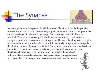
The Synapse
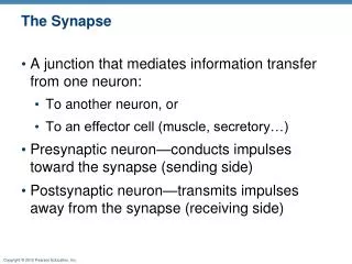
The Synapse. A junction that mediates information transfer from one neuron: To another neuron, or To an effector cell (muscle, secretory…) Presynaptic neuron—conducts impulses toward the synapse (sending side) Postsynaptic neuron—transmits impulses away from the synapse (receiving side).

The Synapse
E N D
Presentation Transcript
The Synapse • A junction that mediates information transfer from one neuron: • To another neuron, or • To an effector cell (muscle, secretory…) • Presynaptic neuron—conducts impulses toward the synapse (sending side) • Postsynaptic neuron—transmits impulses away from the synapse (receiving side)
Types of Synapses • Axodendritic—axon of one to dendrite of another • Axosomatic—axon of one to soma of another • Less common types: • Axoaxonic (axon to axon) • Dendrodendritic (dendrite to dendrite) • Dendrosomatic (dendrite to soma)
Axodendritic synapses Dendrites Axosomatic synapses Cell body Axoaxonic synapses (a) Axon Axon Axosomatic synapses Cell body (soma) of postsynaptic neuron (b) Figure 11.16
Electrical Synapses • Less common than chemical synapses • Neurons are electrically coupled (joined by gap junctions) • Communication is very rapid, and may be unidirectional or bidirectional • Are important in: • Embryonic nervous tissue • Some brain regions
Chemical Synapses • Specialized for the release and reception of neurotransmitters • Typically composed of two parts • Axon terminal of the presynaptic neuron, which contains synaptic vesicles • Receptor region on the postsynaptic neuron
Synaptic Cleft • Fluid-filled space between presynaptic and postsynaptic neurons, found at chemical synapses • Transmission across the synaptic cleft • Is a chemical event (not electrical) • Involves release, diffusion, and binding of neurotransmitters • Provides unidirectional communication between neurons
Synaptic Transmission Summary • Action potential arrives at presynaptic neuron’s axon terminal and opens voltage-gated calcium channels • Calcium enters neuron terminal and causes synaptic vesicle fusion with cell membrane • Neurotransmitter exocytosis occurs • Neurotransmitter diffuses across cleft and binds to receptors on postsynaptic neuron • Ion channels in membrane of post-synaptic cell open, causing excitation or inhibition (graded potential) • Neurotransmitter diffuses away from receptors as it is broken down in the cleft and/or taken back up by pre-synaptic neuron
Chemical synapsestransmit signals fromone neuron to anotherusing neurotransmitters. Presynapticneuron Presynapticneuron Postsynapticneuron 1 Action potentialarrives at axon terminal. 2 Voltage-gated Ca2+channels open and Ca2+enters the axon terminal. Mitochondrion Ca2+ Ca2+ Ca2+ Ca2+ Synapticcleft 3 Ca2+ entry causesneurotransmitter-containing synapticvesicles to release theircontents by exocytosis. Axonterminal Synapticvesicles 4 Neurotransmitterdiffuses across the synapticcleft and binds to specificreceptors on thepostsynaptic membrane. Postsynapticneuron Figure 11.17, step 4
Ion movement Graded potential 5 Binding of neurotransmitteropens ion channels, resulting ingraded potentials. Figure 11.17, step 5
Enzymaticdegradation Reuptake Diffusion awayfrom synapse 6 Neurotransmitter effects are terminatedby reuptake through transport proteins,enzymatic degradation, or diffusion awayfrom the synapse. Figure 11.17, step 6
Termination of Neurotransmitter Effects • Neurotransmitter effect is terminated in a few milliseconds by • Degradation by enzymes • Reuptake by astrocytes or axon terminal • Diffusion away from synaptic cleft
Synaptic Delay • Neurotransmitter must be released, diffuse across synapse, and bind to receptors • Synaptic delay = time needed to do this (0.3-5.0 ms) • Synaptic delay is the rate-limiting step of neural transmission (in short neurons at least)
Postsynaptic Potentials • Graded potentials • Strength determined by: • Amount of neurotransmitter released • Time the neurotransmitter is in the area • Types of postsynaptic potentials • EPSP—excitatory postsynaptic potentials • IPSP—inhibitory postsynaptic potentials
Excitatory Synapses and EPSPs • Neurotransmitter binds to and opens chemically gated channels that allow simultaneous flow of Na+ and K+ in opposite directions • Na+ influx is greater than K+ efflux, causing a net depolarization • EPSP helps trigger AP at axon hillock if EPSP is of threshold strength and opens the voltage-gated channels
An EPSP is a local depolarization of the postsynaptic membrane that brings the neuron closer to AP threshold. Neurotransmitter binding opens chemically gated ion channels, allowing the simultaneous pas- sage of Na+ and K+. Membrane potential (mV) Threshold Stimulus Time (ms) (a) Excitatory postsynaptic potential (EPSP) Figure 11.18a
Inhibitory Synapses and IPSPs • Neurotransmitter binds to and opens channels for K+ or Cl– • Causes hyperpolarization (inside of cell becomes more negative) • Reduces the postsynaptic neuron’s ability to produce an action potential
An IPSP is a local hyperpolarization of the postsynaptic membrane and drives the neuron away from AP threshold. Neurotransmitter binding opens K+ or Cl– channels. Membrane potential (mV) Threshold Stimulus Time (ms) (b) Inhibitory postsynaptic potential (IPSP) Figure 11.18b
Integration: Summation • One EPSP cannot induce an action potential • EPSPs can sum to reach threshold • IPSPs and EPSPs can cancel each other out
Integration: Summation • Temporal summation • One presynaptic neuron sends several or many impulses in a short time to the postsynaptic neuron • Spatial summation • Multiple presynaptic neurons stimulate the post-synaptic neuron simultaneously
E1 E1 Threshold of axon of postsynaptic neuron Resting potential E1 E1 E1 E1 Time Time (a) No summation: 2 stimuli separated in time cause EPSPs that do not add together. (b) Temporal summation: 2 excitatory stimuli close in time cause EPSPs that add together. Excitatory synapse 1 (E1) Excitatory synapse 2 (E2) Inhibitory synapse (I1) Figure 11.19a, b
E1 E1 E2 I1 E1 + E2 I1 E1 + I1 Time Time (c) Spatial summation: 2 simultaneous stimuli at different locations cause EPSPs that add together. (d) Spatial summation of EPSPs and IPSPs: Changes in membane potential can cancel each other out. Figure 11.19c, d
Neurotransmitters • Most neurons make two or more neurotransmitters, which are released at different stimulation frequencies • 50 or more neurotransmitters have been identified • Classified by chemical structure and by function
Chemical Classes of Neurotransmitters • Acetylcholine (Ach) • Released at neuromuscular junctions and some autonomic neurons • Synthesized in the pre-synaptic neuron • Degraded by acetylcholinesterase (AChE)
Chemical Classes of Neurotransmitters • Biogenic amines include: • Norepinephrine (NE) • Epinephrine • Serotonin • Many others • Broadly distributed in the brain • Play roles in emotional behaviors and the biological clock
Chemical Classes of Neurotransmitters • Amino acids include: • GABA—Gamma ()-aminobutyric acid • Glutamate • Many others
Functional Classification of Neurotransmitters • Excitatory (depolarizing) and/or inhibitory (hyperpol.) • Determined by receptor type on postsynaptic neuron • GABA usually inhibitory • Glutamate, epinephrine usually excitatory • Acetylcholine • Excitatory at neuromuscular junctions in skeletal muscle • Inhibitory in cardiac muscle
Neurotransmitter Actions • Direct action • Neurotransmitter binds to channel-linked receptor and opens ion channels • Promotes rapid responses • Examples: ACh; glutamate and GABA at some of their synapses
Neurotransmitter Actions • Indirect action • Neurotransmitter binds to a G protein-linked receptor and acts through an intracellular second messenger • Promotes long-lasting effects • Examples: Norepinephrine, epinephrine, serotonin; glutamate and GABA at some of their synapses