Synapse formation
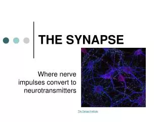
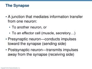
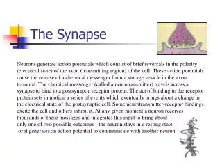
Synapse formation. Raghav Rajan Bio 334 – Neurobiology I September 5th 2013. Synapses are the connections between two neurons – can be electrical or chemical. Typical chemical synapse Presynaptic axon Postsynaptic dendrite Synaptic cleft Since they are small – difficult to visualize

Synapse formation
E N D
Presentation Transcript
Bio 334 - Neurobiology I - Synapse and map formation Synapse formation Raghav Rajan Bio 334 – Neurobiology I September 5th 2013
Bio 334 - Neurobiology I - Synapse and map formation Synapses are the connections between two neurons – can be electrical or chemical Typical chemical synapse Presynaptic axon Postsynaptic dendrite Synaptic cleft Since they are small – difficult to visualize Considerable debate about their presence Synapse – term coined by Charles Sherrington http://en.wikipedia.org/wiki/File:Active_zone3.JPG
Bio 334 - Neurobiology I - Synapse and map formation Synapses evolved about 1.1 million years ago, but some components were present even earlier http://www.lscp.net/persons/ramus/fr/GDP1/papers/verhage00.pdf
Bio 334 - Neurobiology I - Synapse and map formation Complexity of the signalling process in the postsynaptic density has increased greatly http://www.lscp.net/persons/ramus/fr/GDP1/papers/verhage00.pdf
Bio 334 - Neurobiology I - Synapse and map formation Synapse location and number are not random – instead they are regulated Excitatory synapses are typically on spine heads Inhibitory synapses are typically on cell bodies, proximal dendrites or spike necks Synapse number can vary depending on target neurons http://www.richardsmrt.com/?page_id=86
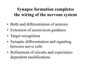
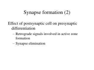
Bio 334 - Neurobiology I - Synapse and map formation Three general observations related to synapse formation Synaptic building blocks are manufactured by neurons even before they make contacts with each other Intercellular signaling, signals from glia, extracellular matrix, neighbouring neurons – all participate in synaptogenesis Synapses mature over the course of development – experience dependent plasticity, critical periods, etc....
Bio 334 - Neurobiology I - Synapse and map formation Study of synapses gained speed in 1950s with the advent of two new techniques Electron microscopy Intracellular recordings http://faculty.washington.edu/chudler/java/em.html http://en.wikipedia.org/wiki/File:RL_Squid_Synapse_2.jpg
Bio 334 - Neurobiology I - Synapse and map formation Pre and post-synaptic membranes come close to each other in a newly formed synapse But, not much can be seen in terms of presynaptic or postsynaptic specializations Difficult to see newly forming synapses – since there is nothing much to see at this stage Dan H Sanes, Thomas A Reh, William A Harris. Development of the Nervous System 2005 – Chapter 8
Bio 334 - Neurobiology I - Synapse and map formation A lot of information about synapse formation comes from watching synapse formation in culture After contact, filopodia retract Slowly pre and post-synaptic parts mature Extracellular matrix also matures Dan H Sanes, Thomas A Reh, William A Harris. Development of the Nervous System 2005 – Chapter 8
Bio 334 - Neurobiology I - Synapse and map formation Important features of synapses - location, location, location – but how is this determined Inputs far away on the dendritic tree have less impact at the cell body Recent studies show that this is not entirely true – may depend on the properties of dendrites in different neurons http://www.sciencedirect.com/science/article/pii/S0960982200000348?np=y
Bio 334 - Neurobiology I - Synapse and map formation First synapses form on growth cones or extremities – later on cell bodies Axo-dendritic synapses onto dendritic growth cones Axo-muscle synapses onto muscle myopodia May even be regulated by glia controlling accessibility to various parts of the post-synaptic cell
Bio 334 - Neurobiology I - Synapse and map formation Pre and post-synaptic structures can form independent of partners Clustering of post-synaptic alpha-2 adrenergic receptors without any presynaptic membrane in rat visual cortex (p4) Presynaptic terminal with vesicles in a Drosophila mutant that does not make muscle Dan H Sanes, Thomas A Reh, William A Harris. Development of the Nervous System 2005 – Chapter 8
Bio 334 - Neurobiology I - Synapse and map formation Synapse number increases after birth Cat visual cortex Neuron density decreases with increased gliogenesis But neuronal processes grow and start making synapses Dan H Sanes, Thomas A Reh, William A Harris. Development of the Nervous System 2005 – Chapter 8
Bio 334 - Neurobiology I - Synapse and map formation Growth cones of axons can release neurotransmitters spontaneously before formation of contacts Dan H Sanes, Thomas A Reh, William A Harris. Development of the Nervous System 2005 – Chapter 8
Bio 334 - Neurobiology I - Synapse and map formation Functional synapses can form very quickly in culture soon after contact Muscle cell brought into contact with neurite Spontaneous currents and evoked currents change rapidly Working synapse is produced quickly But, functional maturation can take days to weeks Dan H Sanes, Thomas A Reh, William A Harris. Development of the Nervous System 2005 – Chapter 8
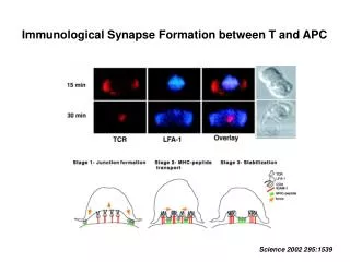
Bio 334 - Neurobiology I - Synapse and map formation Stages in synapse formation – 1 – contact formation – Ca2+, PKC, cAMP all play a role Contact with the correct postsynaptic target can induce a Ca2+ increase in the growth cone triggering cytoskeletal changes Astrocytes also play a role in this process Dan H Sanes, Thomas A Reh, William A Harris. Development of the Nervous System 2005 – Chapter 8
Bio 334 - Neurobiology I - Synapse and map formation Stages in synapse formation – 2 – Increase in adhesion between growth cone and target cell Increase in adhesion between growth cone and target cell within 15 minutes of contact Nectins, cadherins, etc.... Dan H Sanes, Thomas A Reh, William A Harris. Development of the Nervous System 2005 – Chapter 8
Bio 334 - Neurobiology I - Synapse and map formation Stages in synapse formation – 3 – converting sticky growth cone to a presynaptic terminal Presynaptic terminals can mature quickly without concomitant maturation of postsynaptic partners Mature forms of either partner can stimulate maturation of the other Dan H Sanes, Thomas A Reh, William A Harris. Development of the Nervous System 2005 – Chapter 8
Bio 334 - Neurobiology I - Synapse and map formation Signaling pathways again ..... they can change growth cones into presynaptic terminals Different signaling pathways activated by contact stimulate change of growth cone into presynaptic terminal Dan H Sanes, Thomas A Reh, William A Harris. Development of the Nervous System 2005 – Chapter 8
Bio 334 - Neurobiology I - Synapse and map formation Postsynaptic clustering of receptors can be autonomous ACh receptors stained with alpha-bungarotoxin Mouse diaphragm muscle Localization in the centre even in mutants without axon ingrowth Stabilization by presynaptic contact Dan H Sanes, Thomas A Reh, William A Harris. Development of the Nervous System 2005 – Chapter 8
Bio 334 - Neurobiology I - Synapse and map formation Postsynaptic clustering of receptors can also be induced by contact with the right neurons Clustering can be induced by contact Dan H Sanes, Thomas A Reh, William A Harris. Development of the Nervous System 2005 – Chapter 8
Bio 334 - Neurobiology I - Synapse and map formation Agrin, a proteoglycan, is another cluster-inducing molecule In this case, basal lamina also produce agrin and can induce clustering of post-synaptic Ach receptors (frog NMJ) Agrin also produced by motor neurons Dan H Sanes, Thomas A Reh, William A Harris. Development of the Nervous System 2005 – Chapter 8
Bio 334 - Neurobiology I - Synapse and map formation Overall take home of synapse formation Highly specific in terms of location and connections Both sides play a role And there may be other players – glia, extracellular matrix The order of events is not completely understood – may be different for different synapses NOT FIXED – STILL ROOM FOR PLASTICITY