Synapse Formation
Synapse Formation. for questions: contact Kerri at kamassey@ucsd.edu. Amino Acids help determine the final shape and function of proteins By changing the properties of the AAs, you can change how proteins fold or make subtler changes: Change channel kinetics Alter Ion permeability

Synapse Formation
E N D
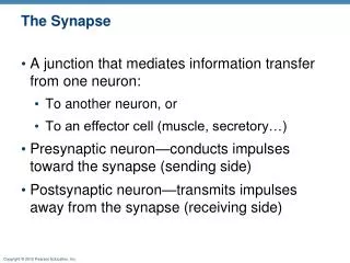
Presentation Transcript
Synapse Formation for questions: contact Kerri at kamassey@ucsd.edu
Amino Acids help determine the final shape and function of proteins • By changing the properties of the AAs, you can change how proteins fold or make subtler changes: • Change channel kinetics • Alter Ion permeability • Alter or Introduce Phosphorylation sites
Culture Systems Brain Slices preserve architecture and glia, can be used to look at more local changes and study electrophysiology Whole brain extracts can be used to study global changes In vivo studies look at the whole animal Dissociated cultures allow individual cells to be studied more easily: A single cell can be followed over space or time with far less background. Explant cultures (organotypic) are “chunks” of tissue that are removed from the brain and grown in culture. They preserve some architecture and support cells. Useful to coculture cells that are not close together or to look at the interaction of tissues from different time points.
mAB: water mAB: fish Merge Caution on Colocalizationor: Kerri’s Pet Peeve By comparing the Actual colocalization to the probability of random colocalization, you can see if colocalization is greater or less than expected. In this case, the actual colocalization is exactly what you would predict from random overlap. Though you have perfect colocalization, you cannot safely say that fish and water colocalize. Real World Example: Some GABA receptors have a large internal pool of receptors so I get a lot of internal staining. When I look at the colocalization of these receptors with GAD (a stain for GABAergic terminals) I see random colocalization: This does not mean that the two are not associated, but it does mean that I can’t use colocalization to prove or disprove that point. Take Home Message: If one of your stains is diffuse, be careful about drawing conclusions about colocalization. probability of fish: area covered by fish/total area= 15% probability of water: area covered by water/total area= 100% probability of colocalization: p(water)*p(fish)=15% probability of colocalization: p(water)*p(fish)=15% actual colocalization: area covered by yellow/total area=15%
More Cautions on Colocalizationor: Guess what Kerri spends her time doing • Just because two things colocalize, it does not mean that they interact. • Bleedthrough: When one color is really bright, in can “bleed through” and be picked up as another color. When comparing two images, look for a bright green dot that isn’t a bright red dot and vice versa. Always be cautious if things look too good. • Some colors, like AMCA, just aren’t picked up by the camera as well. This will make two signals that are actually the same intensity appear to be at different intensities. When looking at colocalization, this can make two things that do partially overlap appear to be only near each other. • Alternatively, if one color has a strong signal, this can make it appear to cover a larger area. This can make two signals that are apposed to each other appear to overlap. This is more of a problem with widefield fluorescent microscopy than two photon fluorescent microscopy.
RNAi • Dicer, an endonuclease specific to double stranded RNA, cleaves large stretches of dsRNA into shorter pieces. • These siRNAs recruit a complex of proteins that includes a distinct endonuclease. • This complex denatures the small siRNA. The antisense strand is used to seek and destroy single stranded RNA in the cell. • This probably developed as an antiviral response. It allows degredation of foreign double stranded RNA as well as destruction of mRNA that is being actively transcribed.
Introducing double stranded RNA into mammalian cells triggers a global halt in protein synthesis which will kill off the cells. • To bypass this, siRNAs are introduced directly. Sequences that are under 30 nucleotides tend to not trigger the interferon response. They are a natural intermediate and the remainder of the process is as described above.
siRNA • siRNAs are most efficient when they are 21-23 nucleotides long with overhanging ends. • They should be directed against a region that doesn’t show much homology with genes you don’t want to interfere with. • They should be directed against a region downstream from the start codon. This will help to avoid the complex being blocked from reaching the mRNA by upstream modulators that bind in the untranslated region. • In mammalian cells, the effect only lasts 4-6 days because the siRNAs eventually are used up or degraded. • To bypass this, scientists are beginning to use replicative-competent viral vectors. • Because of it’s specificity, siRNA may be useful to inhibit translation of mutant gene products when the normal gene product is needed for cell survival. It also has application for cancer and viral infections.