Capture and Detection Antibodies
220 likes | 375 Vues
Capture and Detection Antibodies. HCV ELISA.

Capture and Detection Antibodies
E N D
Presentation Transcript
HCV ELISA • Classical, parentally transmitted non-A, non-B hepatitis has been shown to be due to a small RNA virus, HCV. Non-A, non-B hepatitis represents greater than 90% of transfusion-associated hepatitis cases in the world, and up to 10% of transfusions have been estimated to result in non-A, non-B hepatitis.
This HCV ELISA kit has succeeded in detecting HCV antibodies by using a highly antigenic polyprotein derived from genes encoding essential epitopes sequences of HCV strains prevalent in several regions of the world. It may provide a more comprehensive and effective diagnostic assay to HCV antibodies
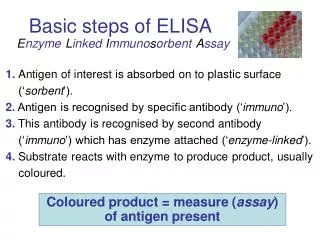
Test Principle • The test principle is based on indirect enzyme immunoassay. • Micro titer wells are coated with certain amount of HCV recombinant antigens including NS3,NS4,NS5 and CORE antigens. • Then serum samples are allowed to react with solid phase antigens. • If HCV-specific antibodies (IgG and IgM) are presented in the serum they will bind to HCV antigens through their individual Fab section. • After incubation, the wells are washed to remove unbound antibodies and anti-human antibodies (IgG/IgM) conjugated with HRP is added into the wells following another incubation and wash step. • A solution of TMB is added and incubated for 15 minutes, resulting in the development of a blue color. The color development is stopped with the addition of stop solution, and the color is changed to yellow and measured spectrophotometricallyat 450 nm. • The concentration of specific anti HCV is directly proportional to the color intensity of the test sample.
Horseradish peroxidase • The enzymehorseradish peroxidase (HRP), found in horseradish, is used extensively in biochemistry applications primarily for its ability to amplify a weak signal and increase detectability of a target molecule.
Applications • Horseradish peroxidase is a 44,173.9dalton glycoprotein with 4 lysine residues for conjugation to a labelled molecule. • It produces a coloured, fluorimetric or luminescent derivative of the labeled molecule allowing it to be detected and quantified. • HRP is often used in conjugates (molecules that have been joined genetically or chemically) to determine the presence of a molecular target. For example, an antibody conjugated to HRP may be used to detect a small amount of a specific protein in a western blot.
Here, the antibody provides the specificity to locate the protein of interest and the HRP enzyme, in the presence of a substrate, produces a detectable signal. • Horseradish peroxidase is also commonly used in techniques such as ELISA and Immunohistochemistry.
Horseradish peroxidase is ideal in many respects for these applications • because it is smaller, more stable and less expensive than other popular alternatives such as alkaline phosphatase. • It allows generation of strong signals in a relatively short time span.
Substrates • Alone, the HRP enzyme, or conjugates there of, is of little value; its presence must be made visible using a substrate that when oxidized by HRP using hydrogen peroxide as the oxidizing agent, yields a characteristic change that is detectable by spectrophotometric methods
Numerous substrates for the horseradish peroxidase enzyme have been described commercialized to exploit the desirable features of HRP. • These substrates fall into several distinct categories. HRP catalyzes the conversion of chromogenic substrates (e.g. TMB, DAB, ABTS) into colored molecules, and produces light when acting on chemiluminescent substrates (e.g. SuperSignal, ECL).
3,3’,5,5’-Tetramethylbenzidine • TMB can act as a hydrogen donor for the reduction of hydrogen peroxide to water by peroxidase enzymes such as horseradish peroxidase. • The resulting diimine causes the solution to take on a blue colour, and this colour change can be read on a spectrophotometer at a wavelength of 650 nm. • The reaction can be halted by addition of acid or another stop reagent. Using sulfuric acid turns TMB yellow. The colour may be read at 450 nm.
Material Safety • TMB should be kept out of direct sunlight as it is photosensitive. • It is not known if TMB is carcinogenic and the evidence is contradictory: • TMB is not mutagenic • On that evidence, it has been used as a replacement for carcinogenic compounds such as benzidine ando-phenylenediamine.
Conjugated protein • conjugated protein is a protein that functions in interaction with other chemical groups attached by covalent bonds or by weak interactions. • Many proteins contain only amino acids and no other chemical groups, and they are called simple proteins. However, other kind of proteins yield, on hydrolysis, some other chemical component in addition to amino acids and they are called conjugated proteins. The nonamino part of a conjugated protein is usually called its prosthetic group. Conjugated proteins are classified on the basis of the chemical nature of their prosthetic groups. • Some examples of conjugated proteins are lipoproteins, glycoproteins, phosphoproteins, hemoproteins, flavoproteins, metalloproteins, phytochromes, cytochromes and opsins.
In chemistry, spectrophotometry is the quantitative measurement of the reflection or transmission properties of a material as a function of wavelength. It is more specific than the general term electromagnetic spectroscopy in that spectrophotometry deals with visible light, near-ultraviolet, and near-infrared, • Spectrophotometry involves the use of a spectrophotometer. A spectrophotometer is a photometer (a device for measuring light intensity) that can measure intensity as a function of the light source wavelength. Important features of spectrophotometers are spectral bandwidth and linear range of absorption measurement.
Spectrophotometers are most commonly used for the measurement of transmittance or reflectance of a solution or transparent material, like polished glass. However they can also be designed to measure the diffusivity on any of the listed light ranges that usually cover around 250nm - 2500nm using different controls and calibrations. • Within these ranges of light, calibrations are needed on the machine using standards that vary in type depending on the wavelength of the photometric determination • The use of spectrophotometers spans various scientific fields, such as physics, chemistry, biochemistry, and molecular biology.
Bovine serum albumin (also known as BSA or "Fraction V") is a serum albumin protein that has numerous biochemical applications including ELISAs(Enzyme-Linked Immunosorbent Assay), immunoblots, and immunohistochemistry. It is also used as a nutrient in cell and microbial culture. In restriction digests, BSA is used to stabilize some enzymes during digestion of DNA and to prevent adhesion of the enzyme to reaction tubes and other vessels. This protein does not affect other enzymes that do not need it for stabilization. BSA is also commonly used to determine the quantity of other proteins, by comparing an unknown quantity of protein to known amounts of BSA. BSA is used because of its stability, its lack of effect in many biochemical reactions, and its low cost since large quantities of it can be readily purified from bovine blood, a byproduct of the cattle industry.
The nickname "Fraction V" refers to albumin being the fifth fraction of the original Edwin Cohn purification methodology that made use of differential solubility characteristics of plasma proteins. By manipulating solvent concentrations, pH, salt levels, and temperature, Cohn was able to pull out successive "fractions" of blood plasma. The process was first commercialized with human albumin for medical use and later adopted for production of BSA.
Sample diluents: A PBS buffer containing 0.02% Tween 20, protein as stabilizer and 0.05% Kathon CG as preservative. 1. PBS: Phosphate buffered saline – provides stable buffered environment to maintain antibody structure
Wash Buffer:(Tween 20) Nonionic detergent – removes non-specifically bound proteins to reduce background and blocks protein binding sites on the polystyrene • Add 60 mL of Wash Buffer (20X) and dilute to a final volume of 1200 mL with distilled or deionized water. Mix thoroughly. If a smaller volume of Wash Buffer is desired, add 1 volume of Wash Buffer (20X) to 19 volumes of distilled or deionized water. Wash Buffer is stable for 1 month at 2-8°C. Mix well before use.
Microplates: Polystyrene – proteins absorb (bind) by hydrophobic bonds to the polystyrene • Secondary antibody: anti-human antibody linked (conjugated) to horseradish peroxidase (HRP) • Enzyme substrate: 3,3’,5,5’ – tetramethylbenzidine (TMB) – a colorless solution that when oxidized by HRP turns yellow. A substrate is a compound or substance that undergoes change. • Substrates bind to active sites on the surface of enzymes and are converted or changed. In ELISA the specific substrate used changes color. • Substrate Solution:chromogen A and chromogen B should be mixed together in equal volumes up to 15 minutes before use. Refer to the table provided for correct amounts of Substrate Solution to prepare. • chromogen A: contains hydrogen peroxide • chromogen B: containestetramethylbenzidine (TMB) • STOP SOLUTION: (2N sulphuric acid solution (H2SO4).