Download
1 / 59
620 likes | 659 Vues
Discover the stages of digestion and the organization of the digestive system, including the roles of teeth, stomach, small intestine, and more anatomical aspects. Explore the crucial digestive glands and processes of digestion.

E N D
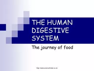
The process of conversion of complex food substances to simple absorbable forms is called digestion and is carried out by our digestive system by mechanical and biochemical methods.
Digestion • Phases Include • Ingestion • Movement • Mechanical and Chemical Digestion • Absorption • Elimination
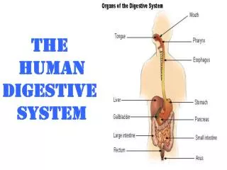
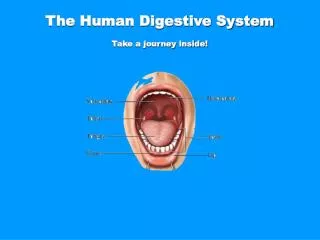
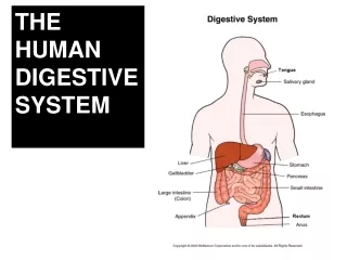
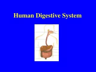
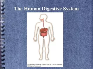
Digestive System Organization • Gastrointestinal (Gl) tract • Tube within a tube • Direct link/path between organs • Structures • Mouth • Pharynx • Esophagus • Stomach • Small intestine • Large Intestine • Rectum
TEETH • An adult human has 32 permanent teeth which are of four different types (Heterodont dentition), namely, incisors (I), canine (C), premolars (PM) and molars (M). • Arrangement of teeth in each half of the upper and lower jaw in the order I, C, PM, M is represented by a dental formula which in human is 2123/2123. • The hard chewing surface of the teeth, made up of enamel, helps in the mastication of food. • The tongue is a freely movable muscular organ attached to the floor of the oral cavity by the frenulum. • The upper surface of the tongue has small projections called papillae, some of which bear taste buds.
Pharynx - serves as a common passage for food & air. A cartilaginous flap called epiglottis prevents entry of food into the glottis-opening of the wind pipe during swallowing. Oesophagus & trachea open into the pharynx • Oesophagus – long , thin tube, passes through neck , thorax , diaphragm &leads to the stomach • Stomach – A muscular sphincter (gastro-oesophageal) regulates the opening of oesophagus into the stomach. • The stomach, located in the upper left portion of the abdominal cavity, has three major parts – a cardiac portion into which the oesophagus opens, a fundic region and a pyloric portion which opens into the first part of small intestine.
Stomach • J-shaped muscular bag that stores the food you eat, breaks it down into tiny pieces. • Mixes food with digestive juices that contain enzymes to break down proteins and lipids. • Acid in the stomach kills bacteria. • Food found in the stomach is called chyme.
Small intestine • Small intestine is distinguishable into three regions, a ‘U’ shaped duodenum, a long coiled middle portion jejunum and a highly coiled ileum. • The opening of the stomach into the duodenum is guarded by the pyloric sphincter. Ileum opens into the large intestine.
Small intestine- has 3 regions, u-shaped duodenum, long coiled middle portion- jejunum, a highly coiled ileum. Opening of the stomach into the duodenum, is guarded by the pyloric sphincter. • Large intestine – consists of – caecum , colon , rectum. Caecum is a small blind sac , which hosts some symbiotic microbes . vermiform appendix – a vestigial organ arises from the caecum. colon has 3 parts- an ascending part, a transverse part & a descending part.
DIGESTIVE SYSTEM The wall of the digestive tube from the mouth to the anus is composed of four basic layers or tunics.
Wall of the alimentary canal • Has 4 layers • Serosa , muscularis , sub- mucosa, mucosa • Serosa - outermost layer, made of thin mesothelium with connective tissues • Muscularis - made of smooth muscles arranged in inner circular & outer longitudinal layer • Submucosa- formed of loose connective tissue containing nerves, blood, lymph vessels • Mucosa- forms irregular folds in the stomach& thin small finger-like foldings called villiin the small intestine . • Villi are richly supplied with blood capillaries. mucosa also forms glands in the stomach & crypts of Lieberkuhn in the intestine
Digestion of Food • BUCCAL CAVITY • performs two major functions, mastication of food and facilitation of swallowing. • The teeth and the tongue with the help of saliva masticate and mix up the food thoroughly. • Mucus in saliva helps in lubricating and adhering the masticated food particles into a bolus. • The bolus is then conveyed into the pharynx and then into the oesophagus by swallowing or deglutition. • The bolus further passes down through the oesophagus by successive waves of muscular contractions called peristalsis.
The gastro-oesophageal sphincter controls the passage of food into the stomach. • The saliva secreted into the oral cavity contains electrolytes (Na+, K+, Cl-, HCO-) and enzymes, salivary amylase and lysozyme. • About 30 per cent of starch is hydrolysed here by this enzyme (optimum pH 6.8) into a disaccharide – maltose. • Lysozyme present in saliva acts as an antibacterial agent that prevents infections.
The mucosa of stomach has gastric glands. Gastric glands have three major types of cells namely - (i) mucus neck cells which secrete mucus; (ii) peptic or chief cells which secrete the proenzymepepsinogen; and (iii) parietal or oxyntic cells which secrete HCl and intrinsic factor (factor essential for absorption of vitamin B12).
DIGESTION IN STOMACH • The stomach stores the food for 4-5 hours. • The food mixes thoroughly with the acidic gastric juice of the stomach by the churning movements of its muscular wall and is called the chyme. • The proenzymepepsinogen, on exposure to hydrochloric acid gets converted into the active enzyme pepsin, the proteolytic enzyme of the stomach.
Pepsin converts proteins into proteoses and peptones (peptides). The mucus and bicarbonates present in the gastric juice play an important role in lubrication and protection of the mucosal epithelium from excoriation by the highly concentrated hydrochloric acid. HCl provides the acidic pH (pH 1.8) optimal for pepsins. Rennin is a proteolytic enzyme found in gastric juice of infants which helps in the digestion of milk proteins. Small amounts of lipases are also secreted by gastric glands.
IN THE SMALL INTESTINE • Digestion is brought about by bile juice , pancreatic juice, intestinal juice. • The pancreatic juice contains inactive enzymes – trypsinogen, chymotrypsinogen, procarboxypeptidases, amylases, lipases and nucleases. • Trypsinogen is activated by an enzyme, enterokinase, secreted by the intestinal mucosa into active trypsin, which in turn activates the other enzymes in the pancreatic juice. • The bile released into the duodenum contains bile pigments (bilirubin and bili-verdin), bile salts, cholesterol and phospholipids but no enzymes. • Bile helps in emulsification of fats, i.e., breaking down of the fats into very small micelles. Bile also activates lipases.
INTESTINAL JUICE OR SUCCUS ENTERICUS. • The intestinal mucosal epithelium has goblet cells which secrete mucus . • The secretions of the brush border cells of the mucosa alongwith the secretions of the goblet cells constitute the intestinal juice or succusentericus. • This juice contains a variety of enzymes like disaccharidases (e.g., maltase), dipeptidases, lipases, nucleosidases, etc.
The mucus along with the bicarbonates from the pancreas protects the intestinal mucosa from acid as well as provide an alkaline medium (pH 7.8) for enzymatic activities. • Sub-mucosal glands (Brunner’s glands) also help in this.
The enzymes in the succusentericusact on the end products of the above reactions to form the respective simple absorbable forms.
Large Intestine • About 5 feet long • Accepts what small intestines don’t absorb • Rectum (short term storage which holds feces before it is expelled).
The breakdown of biomacromolecules mentioned above occurs in the duodenum region of the small intestine. • The simple substances thus formed are absorbed in the jejunum and ileum regions of the small intestine. • The undigested and unabsorbed substances are passed on to the large intestine.
The functions of large intestine are: (i) absorption of some water, minerals and certain drugs; (ii) secretion of mucus which helps in adhering the waste (undigested) particles together and lubricating it for an easy passage.
The undigested, unabsorbed substances called faeces enters into the caecum of the large intestine through ileo-caecal valve, which prevents the back flow of the faecal matter. • It is temporarily stored in the rectum till defaecation.
ABSORPTION OF DIGESTED PRODUCTS • It is carried out by passive, active or facilitated transport mechanisms. • Small amounts of monosacharides like glucose, amino acids and some of electrolytes like chloride ions are generally absorbed by simple diffusion. • The passage of these substances into the blood depends upon the concentration gradients.
However, some of the substances like fructose and some amino acids are absorbed with the help of the carrier ions like Na+. This mechanism is called the facilitated transport. • Transport of water depends upon the osmotic gradient.
Absorption of digested food • It occurs (i) actively, against concentration gradient, with the expenditure of energy (ii) passively by osmosis, facilitated diffusion and simple diffusion. • Glucose, amino acids and Na+ are absorbed actively. • Water- by osmosis • Fructose- facilitated diffusion • Certain small soluble molecules – simple diffusion
Absorption • It occurs within • the ileum in • finger-like • projection • known as • Villi.
Overview of Absorption • Absorptive mechanisms • Passive diffusion • Facilitated diffusion • Active transport
ABSORPTION OF FATTY ACIDS INTESTINAL LUMEN bile salts + MICELLES carbohydrates EMULSIFICATION DROPLETS FAT GLOBULES (triglycerides) free fatty acids, monoglycerides proteins amino acids EPITHELIAL CELL CHYLOMICRONS INTERNAL ENVIRONMENT
Fat absorption • Fatty acids& glycerol being insoluble cannot be absorbed into the blood • They are incorporated into small droplets called micelles which move into the intestinal mucosa • They are then transformed into small protein coated fat globules called chylomicrons which are transported into the lymph vessels • Lymph vessels ultimately release the absorbed substances into the blood stream
Summary of absorption in different parts of digestive system