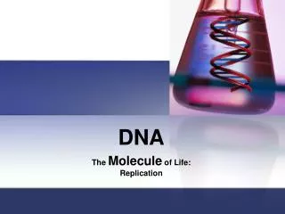
Replication and Cell Division Notes - DNA Structure, Replication Steps, and Mitosis Phases
Explore the fascinating world of DNA structure, replication, and cell division through Maurice Wilkins, Rosalind Franklin, James Watson, and Francis Crick's groundbreaking contributions in 1952-1953. Learn about the double helix shape, nucleotides, nitrogen bases, DNA replication process, mitosis phases, and cell cycle regulation.

Replication and Cell Division Notes - DNA Structure, Replication Steps, and Mitosis Phases
E N D
Presentation Transcript
Replication / Cell Division Notes Chapter 10 and 11
Maurice Wilkins and Rosaline Franklin - 1952 • Photographed DNA using x-rays (crystallography) • Showed a wide, tightly coiled molecule with a helix shape
James Watson and Francis Crick - 1953 • Used Franklin’s DNA x-ray to determine structure of DNA • DNA’s shape • Double helix • 2 strands • Rungs (nitrogen bases)
DNA Structure • Shape – double helix • Location – chromosomes contain DNA • Found in nucleus of eukaryotic cells • Length – long • E. coli – 4,639,221 base pairs or 1.6mm long • 1 m of DNA in each Eukaryotic cell
Nucleotides • Nucleotide consists of: • 5-carbon sugar – deoxyribose • Phosphate group • One of 4 nitrogen bases • Adenine • Thymine • Guanine • Cytosine
Nitrogen Bases • Base pair rule • Adenine always pairs with thymine • A:T • Guanine always pairs with cytosine • G:C • Adenine and guanine are purines • Thymine and cytosine are pyrimidines • Bases are held together by hydrogen bonds (weak)
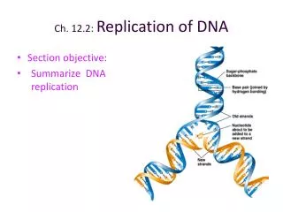
DNA Replication • Process by which DNA is copied • DNA reproduces prior to cell division • Steps • DNA double helix unwinds and unzips • Enzymes break H2 bonds between bases, separating 2 strands • Each parent strand of DNA serves as a template for each new strand • New strands are assembled from free nucleotides • DNA polymerase matches the bases on parent strand creating new double helixes
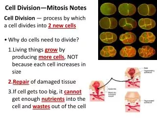
Why Cells Divide • Cell size • Maintain large surface area to volume ratios to transport materials easily • Minimum • DNA • Protein • Internal structures to survive and reproduce • Maximum • Obtain adequate nutrients • Dispose of wastes
Why Cells Divide • Growth • Result of cells producing new cells by cell division • Differentiation – when cells develop into specialized shapes • Repair • Regeneration • Replace dead cells • Skin cells replace every 28 days
Why Cells Divide • Reproduction • Asexual – offspring prod. by 1 parent • Ex. Bacteria, protists, plants and some animals • Cell division (mitosis) replicates chromosomes of one parent cell • Offspring are genetically identical to parent • Gives rise to diploid daughter cells (2N) • Sexual – offspring have combination of genetic material from 2 parents • Cell division (meiosis) results in offspring genetically different from parents • Results in 4 haploid (N) cells
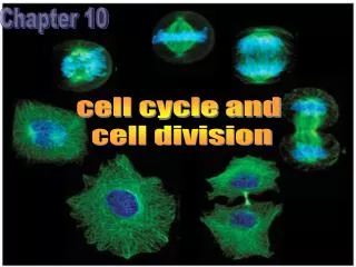
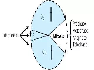
How Do Cells Divide? • Cell cycle – period from beginning of 1 cell division to beginning of the next cell division • 2 parts • Growth • Preparation • Cell divides in 2 stages • Mitosis (nuclear division) • Cytokinesis is (cytoplasm division)
Interphase • Part of cell cycle that occurs between divisions • Longest part of cell cycle – 90% of time • Stages • G1 or gap 1 – 1st stage • Characterized by growth and development • S or synthesis – 2nd stage • Chromosomes in nucleus replicate forming 2 identical structures called sister chromatids • Sister chromatids are visible as cell division begins • Joined together at a point called a centromere • Chromatin – thin, fibrous form of DNA and protein that make up a chromosome
Phases of Mitosis • Prophase – 1st phase • Chromosomes condense and sister chromatids visible • Nuclear membrane and nucleolus break and disappear • Spindle fibers (from cytoskeleton) attach to centromere & begin to move sister chromatids toward center of cell
Phases of Mitosis • Metaphase – 2nd phase • Chromosomes pull to center of cell and line up on the metaphase plate
Phases of Mitosis • Anaphase – 3rd phase • Centromeres divide, spindle fibers pull chromatids apart and towards separate poles (ends) • Once separated, the chromatids are 2 identical sets of daughter chromosomes
Phases of Mitosis • Telophase – 4th phase • 2 daughter nuclei are formed • Nuclear envelope forms around each set of chromosomes • Chromosomes uncoil to form chromatin • Animal cells – each new nucleus has a pair of centrioles outside its nuclear envelope
Phases of Mitosis • Cytokinesis – cytoplasm divides • Begins during telophase as daughter nuclei form • Animal cell • Cell membrane at the center of parent cell folds inward = cleavage furrow • Furrow develops and 2 distinct cells with complete membranes are formed • Plant cell • Membrane fragments fuse to form a cell plate between the 2 new nuclei • A new cell wall forms between the membranes of the cell plate • A complete cell wall that divides the 2 daughter cells is formed
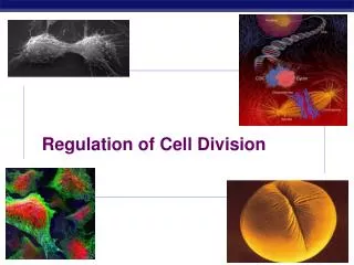
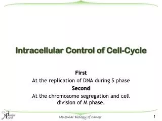
Regulating Cell Cycle • Cyclins • Proteins found in dividing cells • Regulate timing of cell cycle in eukaryotic cells • Figure 10-7 • Internal regulators – proteins that respond to events inside cell • External regulators – proteins that respond to events outside the cell • Cancer cells do not respond to either external or internal signals that regulate the growth of most cells
Meiosis • Before meiosis • A diploid cell replicates its chromosomes • It will now consist of 2 sister chromatids joined by a centromere
Phases of Meiosis • Meiosis I • Prophase I – each homologous pair of chromosomes attach to one another and equals 4 sister chromatids called tetrad • Metaphase I – chromosomes move to the center of the cells • Anaphase I – chromosomes are pulled to the poles • Telophase I – 2 daughter cells are formed that are haploid and consist of 2 sister chromatids • Each daughter cell now undergoes Meiosis II
Phases of Meiosis • Meiosis II • Prophase II – sister chromatids begin to move to the center of the cell • Metaphase II – centromeres split in middle of cell • Anaphase II – individual chromosomes are pulled to the poles • Telophase II – 4 haploid daughter cells result • (2 from each Meiosis I daughter cell)
Phases of Meiosis • Cytokenisis • In human males • All 4 haploid nuclei form sperm • In human females • Only 1 haploid nuclei forms an egg • The other 3 nuclei receive almost no cytoplasm and do not form gametes
Regulating the Cell Cycle • Proteins found in dividing cells • Regulate timing of cell cycle in eukaryotic cells • Internal regulators – proteins respond to events inside • External regulators – proteins respond to events outside cell • CDKs • Involved in regulation of transcription of mRNA • Target for anti-cancer treatment
Genes and Cancer • Causes of cancer • Mutations that change the genes that control cell growth and specializations • Can be inherited • Environmental factors • Combination of genetic and envir.
Oncogene • A gene that causes a cell to become cancerous • Caused by a mutation in a growth factor gene • Caused by a DNA replication error • Multiple copies of a single growth factor gene • Caused by a change in a gene’s location
Environmental Factors • Mutagens – factor in the envir. that can cause mutations • Radiation • Chemicals (tobacco prod.) • Carcinogen – agent that causes or tends to cause cancer by replacing or changing DNA • Tars • Chemicals in smoked meat • Viruses • Radiation (UV – sunlight) • Drugs • Coal tars (hair dyes)
Karyotype • Picture of homologous pairs of chromosomes • Can show a various abnormalities • Nondisjunction – when chromosomes fail to separate during meiosis • Can result in too few or too many chromosomes