Download
1 / 23
260 likes | 512 Vues
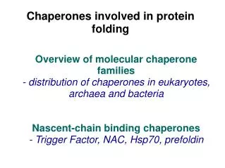
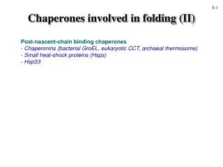
Chaperones involved in folding (II). 8-1. Post-nascent-chain binding chaperones Chaperonins (bacterial GroEL, eukaryotic CCT, archaeal thermosome) Small heat-shock proteins (Hsps) Hsp33. 8-2. alpha-beta hemoglobin heterodimer. a. A chaperone for a -hemoglobin.

E N D
Chaperones involved in folding (II) 8-1 • Post-nascent-chain binding chaperones • Chaperonins (bacterial GroEL, eukaryotic CCT, archaeal thermosome) • Small heat-shock proteins (Hsps) • Hsp33
8-2 alpha-beta hemoglobin heterodimer a A chaperone for a-hemoglobin alpha-hemoglobin stabilizing protein (AHSP) b
GroEL/GroES chaperonin system 8-3 • GroEL forms homo-oligomeric toroidal complex dependent on GroES cofactor for function; GroEL is essential for cell viability • GroEL/GroES system may bind 10% of all bacterial cytosolic proteins but recent study shows only a portion of those are completely chaperonin-dependent • Belongs to so-called Group I chaperonins which includes evolutionarily-related bacterial GroEL, mitochondrial Hsp60, and chloroplast Rubisco subunit-binding protein (Rubisco is most abundant protein on earth and requires chaperonin for folding) • Functional mechanism is the best understood of all chaperonins
GroEL/GroES structure 8-4 crystal structure of E. coli GroEL/GroES • GroEL has two stacked heptameric rings (equatorial domains form inter-ring contacts) • GroES forms a single heptameric ring that binds co-axially to one GroEL ring (caps GroEL, preventing polypeptide exit or entry); binds only when GroEL in ATP state • crystals structure without GroES has been solved, and with ATP-gamma S (non-hydrolyzable ATP analogue) • mitochondrial chaperonin (Hsp60) is single-ring; GroES from chloroplasts consists of a fused dimer
GroEL subunit structure 8-5 • chaperonins have 3 domains • equatorial domain is the ATPase • intermediate domain is a flexible hinge; binding of ATP and GroES causes the apical domain to move upward and turn about 90° to the side • apical domain is the polypeptide binding domain; the binding site consists mostly of large, bulky hydrophobic residues (determined by mutation analysis) • GroES binds to the polypeptide binding site; displaces substrate into the cavity
Group I chaperonin:functional cycle 8-6 • large conformational changes occur upon ATP and GroES binding: cavity interior expands ~2 fold, hydrophobic residues in apical domain turn away from the binding site and the interior becomes hydrophilic • ATP --> ADP transition is when folding takes place in the cavity; when ATP is hydrolyzed, and ATP/GroES binds to trans ring (opposite the cis ring), GroES on cis ring dissociates and the polypeptide exits • the polypeptide may not be folded upon exiting; it could undergo another round of folding by either the same chaperonin, another chaperonin, or another chaperone
GroEL mechanism of action 8-7 1. Multivalent binding of substrate 2. Unfolding of substrate (controversial) - evidence that non-native protein is unfolded further upon binding to GroEL and hydrolysis of ATP 3. Combination of multivalent binding, unfolding may re-direct folding intermediates to proper folding pathway once inside hydrophilic chaperonin cavity 4. Infinite dilution??? (‘cage’ model) Paper presentation (next 3 slides): Farr et al. (2000) Multivalent binding of nonnative substrate proteins by the chaperonin GroEL. Cell100, 561-573.
GroEL function: single polypeptide 8-8 • N- and C-termini of GroEL (chaperonins in general) are buried inside the cavity • construct is a fusion between all 7 subunits--protein size is 400 kDa! • the fusion protein assembles properly as judged by em reconstructions • powerful tool for analyzing contribution of individual subunits to binding, etc.
GroEL function: in vivo 8-9 • strain with wild-type GroEL under control of lac promoter (inducible with IPTG) • without IPTG, strain growth arrests • growth restored when covalent GroEL (fusion construct) is present; this represents a growth of ‘++++’ • other constructs were tested in the absence of IPTG; ‘o’ represents no growth, ‘+’ represents very slow growth
GroEL function: in vitro 8-10 • found that covalent GroEL was a bit less active at binding non-native proteins compared to wild-type GroEL; mild protease treatment restored binding • experiment: binding of denatured protein to various constructs, isolation by SEC, and amount of bound proteins quantitated conclusions: > require at least two or three GroEL subunits for binding non-native proteins; these should preferably be in positions 1-3 or 1-4 (i.e., not immediately adjacent) > ability of GroEL/GroES to fold substrate followed similar pattern (not shown)
Group II chaperonin system eukaryal • Group II chaperonins from the eukaryotic cytosol and archaeal cytosol are more closely related to each other than they are to Group I chaperonins • eukaryotic cytosolic chaperonin is called CCT or TRiC, for “Chaperonin containing TCP-1” or “TCP-1 Ring Complex”. TCP-1 was the first subunit of CCT to be characterized. It was found to be present within a hetero-oligomeric complex that contained 8 different (related) chaperonin subunits • 8-fold symmetry (different than GroEL’s 7-fold) • duplication of chaperonin subunits occurred early during evolution (2 billion years ago), as all eukaryotes contain the same 8 orthologues • involved in actin and tubulin biogenesis BUT folds a number of other proteins, e.g., VHL tumour suppressor, myosin, cyclin E, viral capsid, etc. and binds up to 10% of all cellular proteins archaeal • the archaeal chaperonin, termed “thermosome”, consists of 1-3 different subunits, depending on the archaeal lineage • 8- or 9-fold symmetry • function in protein folding; during cellular stresses (>70% cellular protein!) 8-11a
Group II chaperonin structure 8-11b alpha-helical protrusion side view of top ring GroES apical domain apical domain side view of bottom ring intermediate domain intermediate domain thermosome side view equatorial domain equatorial domain GroEL thermosome comparison of GroEL/ES complex (one subunit of GroEL, one subunit of GroES) with single thermosome (alpha) subunit 8 subunits per ring; 4 alpha, 4 beta subunits thermosome top view
Group II chaperonin:functional cycle 8-12 • open or closed states of thermosome (archaeal chaperonin related to CCT) were determined by SAXS experiments in the presence of nucleotides (ADP, ATP) or ADP in the presence of inorganic phosphate (PO4, or Pi) to simulate ADP*Pi transition state • none of the studies have been carried out in presence of substrates; assume ‘open’ conformations can interact with substrate and ‘closed’ state is involved in folding • ATPADP transition somehow causes large conformational change
MODEL WHICH INCORPORATES THE SUBSTRATE Douglas et al. Cell 2011
CCT-actin em reconstruction 8-13 • actin is composed of 4 subdomains, Sub1-Sub4 • hinge between domains Sub3-Sub4 and Sub1-Sub2 is flexible • ATP binds in cleft between large and small domains • actin cannot fold properly in the absense of ATP • CCT-tubulin reconstruction also done; tubulin makes more contacts with CCT subunits
Evolution of chaperonins, prefoldin and actin/tubulin 8-14 • FtsA, actin homologue • FtsZ, tubulin homologue Evolution of eukaryotes • CCT and prefoldin co-evolved; essential for actin/tubulin biogenesis • actin and tubulin are essential components of cytoskeleton • cytoskeleton is required for large number of cell processes unique to eukaryotes, including intracellular movements, engulfment, etc. etc. • hypothesis: eukaryotes could not have evolved without CCT and prefoldin
Small heat-shock proteins 8-15 • found in all three domains of life, usually in multiple copies • form large molecular weight complexes • consist of three distinct domains • can efficiently bind proteins on the aggregation pathway • play important role in thermotolerance; protecting proteins from aggregating under stress conditions • cooperate with other chaperones (e.g., Hsp70) to renature proteins; function, like that of prefoldin, is ATP-independent
Small Hsp crystal structure 8-16 - sizes of small Hsps range from 150 kDa to 800 kDa - smallest functional small Hsp is a nonamer (trimer of trimer) • crystal structure from Methanococcus jannaschii Hsp16 small Hsp (first archaeal genome to be sequenced) (wheat and ? Structures now also known) • spherical shell composed of 24 subunits • 2-, 3-, and 4-fold symmetry • N-terminal domain (first 33 amino acids) were not resolved in the crystal structure; these are likely to be flexible or disordered
Small Hsp surface view 8-17 • immunoglobulin domain fold (same as PapD/ FimC) • dimer interface most extensive (building block) • C-terminal region is exposed on surface • N-terminal region faces interior of the oligomer (N-terminal region was not resolved in the crystal structure)
8-18 Wheat small HSP End view Side view Dodecameric structure van Montfort et al. Nature Structural Biology (2001)
Hsp33: the redox chaperone 8-19 • exclusively bacterial; induced during oxidizing (stress) conditions in the cell Hsp33 oxidizing conditions (e.g., H2O2) Hsp33/Hsp33 dimer Hsp33 • domain-swapped dimer (active form); inactive monomer • activation dependent on redox condition in cell; under oxidizing (stress) conditions, disulfide bridges are formed and dimerization takes place; conserved cysteines • Hsp33 efficient in preventing protein aggregation in vitro Jakob et al. (1999) Cell96, 341.
Hsp33 substrate binding site 8-20 • two possible binding sites that are only available upon dimerization • residues shown are highly conserved across bacterial Hsp33 proteins • ‘multivalent’ binding—again?