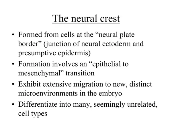
Chapter 13- The neural crest
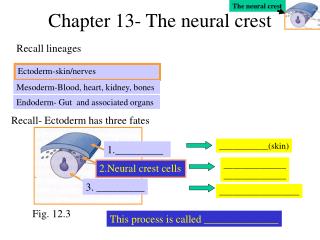
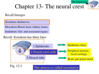
The neural crest. Chapter 13- The neural crest. Recall lineages. Ectoderm-skin/nerves. Mesoderm-Blood, heart, kidney, bones. Endoderm- Gut and associated organs. Recall- Ectoderm has three fates. Epidermis (skin). 1.Epidermis. Peripheral neurons, facial cartilage. 2.Neural crest cells.

Chapter 13- The neural crest
E N D
Presentation Transcript
The neural crest Chapter 13- The neural crest Recall lineages Ectoderm-skin/nerves Mesoderm-Blood, heart, kidney, bones Endoderm- Gut and associated organs Recall- Ectoderm has three fates Epidermis (skin) 1.Epidermis Peripheral neurons, facial cartilage 2.Neural crest cells 3.Neural tube Brain and spinal chord Fig. 12.3 This process is called neurulation
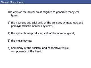
The neural crest Neural crest cell fate depends largely on where they migrate The neural crest is a transient structure Potential cell fates include- 1. neurons and glia 2. medulla of adrenal gland (produces epinephrine) 3. Pigment cells of epidermis 4. Skeletal/connective tissue of head Neural crest- four functional domains 1. Cranial- cartiledge, bone, neurons, glia of face 2. Cardiac 3. Vagal- parasympathetic ganglia 4. Trunk- melanocytes (produce pigment); sensory and sympathetic neurons, medulla Fig. 13.1
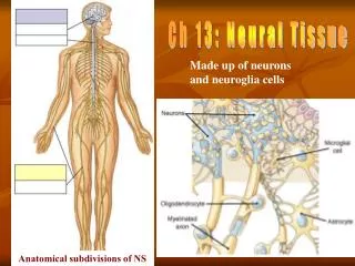
Sensory pathways- conduct info to brain-, spinal cord A quick review of nerve nomenclature 1. Autonomic nervous system • “involuntary controlled muscles”- • CNS sends signals to smooth muscles of heart, blood vessels, iris, pancreas liver, digestive tract, kidney • 1.Parasympathetic- -homeostasis of body systems, originate from hindbrain • 2. Sympathetic-fright and flight reactions- originate form spinal chord 2. Somatic nervous system- -“voluntary controlled organs”- - CNS sends signals to striated muscles communication between various parts of the body (e.g. thallumus, cerebellum) with muscles Figure not in text
The neural crest Path 1-cells travel under epidermis, become melanocytes, colonize hair and skin follicles Epidermis Neural tube Sclerotome Notochord This is a somite A. Start with the Trunk Neural crest Two major paths taken Fig. 13.2 Path 2-cells to side of neural tube and through anterior sclerotome to become sympathetic and sensory neurons Note – Sclerotome will become vertebral cartilage
The neural crest Neural Crest cells Ephrinin sclerotome How do these neural crest cells know where to migrate? 1. Epidermis secrete BMP-4 and BMP-7 - BMP-4 and –7 induce neural crest cells to produce slug and RhoB - Slug dissociates cell-cell tight junctions 2. N- cadherin expression is also lost then regained once reaching final destination 3. Ephrin proteins in extracellular matrix guide cells • Neural crest cells have Eph receptors • Trunk sclerotome express Eph ligand • Binding of Eph receptor to Eph ligand interferes with migration • Thus, Eph proteins tell neural crest cells where not to go Fig. 13.4 4. Stem cell factor allows continued proliferation 5. Other chemotactic and maintenance factors
The neural crest Mash-1 Sympathetic and parasympathetic neurons Neurogenin Sensory neuron • Trunk neural crest cells arepluipotent(can become many cell types) • However, it may be that only certain populations of cells are pluripotent • Some transcription factors have been identified that dictate cell fate Trunk neural crest cell Final cell fate is determined by final environment Fig. 13.6- Fate of a trunk neural crest cell is influenced by FGF2 and glucocorticoids
The neural crest Rhombomeres B. The Cranial neural crest Like the trunk neural crest cells, these can produce glia, neurons and melanocytes But, only cranial neural crest cells can produce cartilage and bone • Recall – the neural tube subdives into forebrain, midbrain and hindbrain • The hind brain then further subdivides into rhombomeres • Each rhombomere is a territory, each produces ganglia, but each has a distinct fate • Rhombomeres sit behind the pharyngeal arches Pharyngeal arches Fig. 13.1
The neural crest Three paths for cranial neural crest cells Rhombomeres in hind brain of neural tube Pharyngeal arches 1. Rhombomere 1+2- to 1st Ph. Arch 3. Rhombo. 6 to 3rd and 4th Ph. Arch 1 2. Rhombo. 4- to 2nd Ph. Arch 2 3 4 Fig. 13.7 Rhombomeres 3 and 5 do not migrate through arches Fate map of pharyngial arches contributions to face formation
The neural crest + retinoic acid No ear WT Fig. 13.8 What determines distinct fates of cranial neural crest cells? Answer- The combination of hox genes Evidence 1. Hoxa-2 KO- neural crest cells of 2nd Ph. Arch transformed into 1st Ph. Arch structures 2. Hoxa-1 and Hoxb-1 double KO- no rhombomere 4 migration 3. Retinoic acid induces more anterior expression of certain Hox genes- - rhombomeres 2 and 3 assume role of rhombomeres 4 and 5
How is neuronal diversity achieved?? 5 ways- 1. Blocking BMP signal allows formation of dorsal neural tube (recall chapter 12) 2. Notch-delta specifies neural fate (not epidermal or glial) 3. Initial location determines neuronal type 4. Migration route further dictates specificity 5. Specific connection made with target organs or other neurons 3 parts described 1. Pathway selection- axons travel along a given route 2. Target selection- axons reach a target, then bind to specific cells 3. Address selection- axons now refine interactions- bind to only a subset of possible targets
A. Hypotheses for pathway selection- • Cell adhesions- Growth cone can adhere to certain cells, but not • others • Laminin – a glycoprotein which appears to pave the road for several axonal migrations • N-CAM 2. Physical barriers- Growth cone can adhere to certain cells, but not others 3. Labeled pathway hypothesis- in insects, a neuron can precisely follow the path of a prior neuron Kallmann syndrome- an infertile man with lack of smell Reason- a single protien directs migration of both olfactory axons and hormone-secreting nerve cells • 4. Repulsion- • Ephrin (recall Fig 13.4) – Growth cones contain Eph receptors- binding prevents migration into undesirable areas • Semaphorin proteins- important in directing axon turns
Fig. 13.20 Rat dorsal spine explant Neuron Outgrowth Neutrin producing cell Fig. 13.21 Sensory Neuron Motor Neuron Unc-6 -/- WT Hypotheses for pathway selection- (Cont.) 5. Diffusible molecules- a. Netrin-1 and –2 are chemotactic • Netrins are homologues of the UNC-6 protein in C. Elegans Loss of Unc-6 prevents migration of both sensory (to ventral) and motor (to dorsal) neurons b. Slit proteins are repulsive
0 min 2 min 6 min 10 min B. Hypotheses for target selection- Target cells secrete short-range chemotactic or chemorepulsive factors Example- NT-3 attracts axons C. Hypotheses for address selection- Fig. 13.24 Growth cone makes contact with a cell, acetylcholine receptors cluster on target cell surface, and a synapse is formed Additional axons synapse target cell, but eventually only one/cell remains Fig. 13.25