
neural crest
E N D
Presentation Transcript
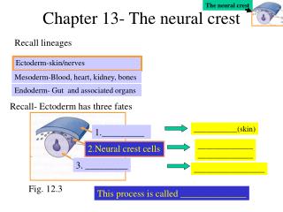
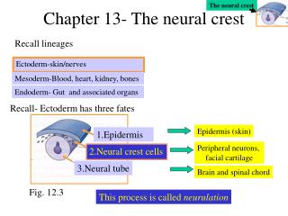
The neural crest • Formed from cells at the “neural plate border” (junction of neural ectoderm and presumptive epidermis) • Formation involves an “epithelial to mesenchymal” transition • Exhibit extensive migration to new, distinct microenvironments in the embryo • Differentiate into many, seemingly unrelated, cell types
Five classes of neural crest cells • Cranial neural crest • Cardiac neural crest • Vagal neural crest • Trunk neural crest • Sacral neural crest Anterior I I I I V Posterior
Neural crest migration pathwaysFigure 13.1 of Gilbert, p.412
Cranial neural crest • Migrate “dorsolaterally” and are precursors of facial structures • Craniofacial cartilage and bones • Cranial neurons and glia • Connective tissue of the face • Jaw and tooth precursors • Bones of the middle ear • More about this class later on…
Cardiac neural crest • Originates in the region between the cranial and trunk neural crest (somites 1-3 in chick) • Develop into melanocytes, neurons, cartilage, connective tissue • Importantly, these cells give rise to the entire musculoconnective tissue wall of the arteries arising from the heart and the septum that separates pulmonary circulation from the aortic circulation • A bit more to come on this class later on…
Vagal/Sacral neural crest • Vagal: • Generate the neurons of the gut • Emigrate from the neural tube near somites 1-7 of the chick • Sacral: • Form posterior to somite 28 of the chick • Differentiate into the neurons of the gut (i.e. those involved in peristalsis)
Trunk neural crest • Migrate along two distinct pathways that lead to different developmental fates • Pathway 1- dorsolateral from neural tube into the ectoderm and continuing to the ventral midline of the embryo. These cells differentiate into melanocytes (pigment cells) • Pathway 2- Ventrolateral migration through the anterior sclerotomes. These cells form dorsal root/sensory ganglia, sympathetic neurons, adrenal medulla and Schwann cells
Aspects of neural crest biology • Specification of the neural crest fate • Initiation of migration (i.e. epithelial to mesenchymal transition) • Guidance to migration route and destination • Final differentiation of neural crest in response to signals present at destination
Neural crest specification • Interactions between the neural plate and the non-neural ectoderm • Juxtaposition of the two tissue types in vitro is sufficient to induce neural crest formation • Neural crest precursors come from both the neural plate AND the prospective epidermis • Non-axial mesodermal signals play an important role in vivo
Signals implicated in neural crest specification in vivo • BMPs - By late gastrulation, BMP4 is expressed very highly at the neural plate border. This expression continues in the dorsal neural tube (as it closes up) as nearby epidermis loses expression of BMP4 • FGFs and Wnts - presumed to come from the underlying mesoderm
Signals specifying neural crest fateFigure 2 from LaBonne and Bronner-Fraser (1999) Annu. Rev. Cell Dev. Biol. 15:81-112
Markers of prospective neural crest cells • Snail and Slug - are Zinc finger transcription factors • Xenopus: snail and slug are expressed both in premigratory and migratory neural crest • Mouse: slug is only expressed in migrating neural crest. Interfering with slug expression can prevent neural crest emigration • Slug knockout mice display no obvious neural crest defects
Markers of neural crest, continued • Pax-3 and Pax-7 - Knockouts mice of each molecule display neural crest defects • Zic-1, Zic-2, Zic-3 - Zinc finger transcription factor homologous to Drosophila pair-rule gene (odd-paired) • Msx-1 - Homeodomain transcription factor. Mouse knockouts have deficiencies in neural crest derivatives
Initiation of neural crest migration • Epithelial to mesenchymal transition occurs prior to migration • Involves changes in cell adhesion molecules • Cadherin switching • Pre-migration expression of N-cadherin and Cadherin 6B • Post-migration expression of Cadherins 7 and 11
Signaling molecule involvement • Rho-family GTP-ases • Implicated in cytoskeletal rearrangements that drive changes in cell behavior • Rho-B is expressed in the dorsal neural tube • C3-exotoxin prevents neural crest cells from separating from the neural tube • RhoB is upregulated by BMP treatment
Factors determining the route of neural crest cell migration • Extracellular matrix (ECM) proteins • Promote migration: fibronectin, laminin, tenascin, collagens, proteoglycans • Prevent migration: Ephrin proteins (!) • Eph receptors on neural crest • Tyrosine kinase domain • Thought to phosphorylate proteins that interfere with the actin cytoskeletal rearrangements • See website 13.1 which discusses this issue
Fibronectin interacts with integrin cell adhesion moleculesFigure 6.34 of Gilbert, p. 171
Negative signals from EphrinsFigure 13.4 of Gilbert, p. 415 Migrating neural crest cells only go where Ephrins are NOT
Key Concepts to this point… • Classes of neural crest and their progeny • Molecules involved in the formation and mirgration of neural crest • Epithelial to mesenchymal transition • Molecular guidance cues • Next topic: Neural crest cell differentiation
Neural crest cell differentiation • Individual neural crest cells are multipotent • Capable of limited self-renewal in an undifferentiated state (like a stem cell) • Usually adopt fates determined by their axial level (anterior-posterior) • Are capable of adopting other fates when transplanted to different axial regions • Exception” the ability to give rise to cartilage seems to only be a trick of the cranial crest • Fate is determined by signals at the destination of each cell
Potential vs. FateFigure 9.28 From Wolpert (1998) Principles of Development, Oxford U. Press
Developmental pathwaysFigure 9.27 From Wolpert (1998) Principles of Development, Oxford U. Press
Signals that differentiate trunk neural crest cellsFigure 3 from LaBonne and Bronner-Fraser (1999) Annu. Rev. Cell Dev. Biol. 15:81-112
Differentiation of melanocytesFigure 9.30 From Wolpert (1998) Principles of Development, Oxford U. Press
Cranial neural crest movement into pharyngeal arches/pouches • Pharyngeal arches/pouches are the anterior-most descendents of the endoderm • Migrate from the anterior hindbrain (rhombomeres 1-6) into the pharyngeal/branchial arches and pouches • Rhombomeres 1&2 ---> 1st pharyngeal arch • Rhombomere 4 ---> 2nd pharyngeal arch • Rhombomere 6 --> 3rd & 4th pharyngeal arches and pouch • Rhombomeres 3&5 send their neural crest into the migrating streams on either side
Some products of the pharyngeal arches and pouches • Arch 1 - Jawbones, inner ear bones, frontonasal processes • Arch 2 - neck cartilage • Arches and pouches 3 and 4 - Thymus, parathyroid gland and thyroid gland • Pharyngeal pouches form between the pharyngeal arches
Cranial neural crest differentiationFigure 13.7 of Gilbert, p. 418
Hox genes specify neural crest fate • HoxA2 knockout - neural crest of 2nd arch become structures characteristic of Arch 1 • HoxA3 knockout - malformed thymus, thyroid, parathyroid glands, neck vertebrae • HoxA1/HoxB1 double knockout - no migration from rhombomere 4 into the 2nd pharyngeal pouch • Retinoic acid causes rhombomeres 2 and 3 derived neural crest to form structures characteristic of rhombomeres 4 and 5
Neural crest and evolution • Neural crest cells are an important unit of the evolution of craniofacial structures • Correlated progression: Changes in one part of the embryo induce changes in another • Neural crest cells from rhombomere 4 form skeletal elements associated with a jaw, the sites of attachment for jaw muscles, the muscles themselves and informs the placement of the proper nerve axon
Cardiac neural crest • Migration occurs from rhombomere 7 into pharyngeal arches 3, 4 and 6 • Fate is determined early. No other neural crest can substitute for it • Express Pax-3, which when knocked out causes heart defects in mice (and defects of thymus, thyroid and parathyroid glands)
Importance of cardiac neural crestFigure 13.10 of Gilbert, p. 423
More Key Concepts • Neural Crest cell differentiation • Location, location, location (and a few paracrine factors) • Interconnections between developmental events over time (connect events across different lectures….) • Dynamic BMP expression levels • Connections between molecular aspects of the development of neighboring tissues