Knee Dislocation and Multiligamentous Injury
1.8k likes | 3.69k Vues
Knee Dislocation and Multiligamentous Injury. Utku Kandemir, MD Revised October 2011 Original Author: William R. Creevy, MS, MD; Mark A. Neault, MD & Brian D. Busconi, MD; March 2004; Robert P. Dunbar, Jr., MD; Revised January 2007. Anatomy: Tibiofemoral Joint. Bones femoral condyles

Knee Dislocation and Multiligamentous Injury
E N D
Presentation Transcript
Knee Dislocation and Multiligamentous Injury Utku Kandemir, MDRevised October 2011Original Author: William R. Creevy, MS, MD; Mark A. Neault, MD & Brian D. Busconi, MD; March 2004;Robert P. Dunbar, Jr., MD; Revised January 2007
Anatomy: Tibiofemoral Joint • Bones • femoral condyles • tibial plateau • Dissimilar surfaces • Little/No inherent bony stability • May be cause of additional instability if fractured
Stabilizers of the Tibiofemoral Joint • Soft tissues: stabilize while allowing ROM • Ligaments • Joint capsule • Menisci • Musculotendinous units (DYNAMIC)
Anatomy – 4 groups of ligaments • ACL • PCL • MCL, posteromedial capsule • LCL & PLC (popliteofibular ligament, popliteus, capsule, ITB, biceps femoris)
Vascular Anatomy • Popliteal artery at risk for being tethered • Adductor hiatus • Soleus arch • If blood flow through popliteal artery disrupted Inadequate blood supply distally
Anatomy: Nerves • Influences LongTerm Outcome • Peroneal nerve • More commonly injured • Tethered around the fibular neck • Mechanism of injury • Tension (Varus ± hyperextension, Translation (Anterior /Posterior dislocation) • Direct impact • Iatrogenic (aggressive varus/hyperextension during EUA (!) • Tibial nerve
Knee Dislocation–Multiligamentous Injury • Disruption of normal relationship of tibiofemoral joint • Usually requires the injury to 2 of the 4 major groups of ligaments
Knee Dislocation–Multiligamentous Injury • Large Spectrum of injury • Increased severity with more structures involved
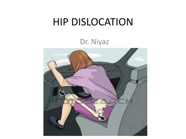
Pathomechanics • May occur not only with high energy but also with low energy • Low energy • Athletic activity (more with contact sports) • Fall down stairs • Jump of the low height • High energy • MVA • PVA • Fall from height
Pathomechanics • Cadaveric study • Progressive Hyperextension model Anterior dislocation • @≈30 degrees: tear of posterior CAPSULE • Followed by tear of PCL & ACL DISLOCATION • @≈50 degrees: rupture of POPLITEAL ARTERY Kennedy JC. 1963
Pathomechanics • Cadaveric study • Combined cruciate ligaments injury in hyperextension • Low rate of strain (100%/sec) midsubstance tear of PCL • High high rate of strain (400%/sec) avulsion of PCL from femur • ACL: Mixed pattern of injury Schenck RC et al. 1999
Why Important? • …serious injury which can have long-term adverse effects which may impair the patient’s return to physical employment and recreational activity. Robertson A et al. 2006
Epidemiology “It is unlikely that any single physician personally cares for more than a few knee dislocations in a lifetime of practice” Meyers MH, Harvey JP. 1971
Epidemiology • True incidence is underreported • Spontaneous reduction • Definition (documented complete dislocation vs. ≥1 cruciate + one collateral injury) • Obesity interferes with exam and mechanism • Presented in a variety of clinical practices • Trauma Center • Sport Medicine • General Orthopaedics
Epidemiology • 0.2 % of all orthopaedic injuries • Young ♂ • MVA, sports trauma • 14-44 % associated w multiple trauma • Bilateral 5 % Robertson A et al. 2006
Ligamentous Injury in Polytrauma Patient • Suspect & Examine in any • Lower extremity long bone fracture • Polytrauma • Head injury • Isolated femoral shaft fx • Associated knee ligament injury: 33% (Walling AK 1982) • Isolated tibial shaft fx • Associated knee ligament injury: 22% (Templeman DC 1989) • Ipsilateral Femoral & tibial shaft fx • Associated knee ligament injury: 32-53% (Szalay MJ 1990, vanRaay JJ 1991)
Diagnosis • If any of the following present r/o Multiligamentous injury (Spontaneous reduction UNDERDIAGNOSED) • Hyperextension • Popliteal ecchymosis • Vascular insufficiency • Peroneal nerve deficit • Diffuse tenderness but Absence of hemartrosis (capsular disruption) • Obese pt low energy fall
Physical Examination • Evaluate soft tissues • Open • Puckering (irreducible dislocation)
Vascular Examination • Color, temperature, Pulses • Dorsalis Pedis a. & Tibialis Posterior a. • ABI (Ankle Brachial Index) • ≥0.9: Serial examination • <0.9: further study/exploration • Johanson, K, JT • Reduce if dislocated and Reexamine
Vascular Examination • ABI ≥0.9 & no signs of vascular injury: Arterial study may not be necessary if • Serial examination q 2-4 hrs for 48 hrs can reliably be performed • If not, arterial study may be ordered to r/o vascular injury Mills WJ 2004, Stannard JP 2004
Vascular Examination • ABI <0.9 OR • Temperature, Color, OR • Expanding swelling (hematoma) around the knee • Arterial study • Arteriography in OR ( on table by surgeon) • Angiography (radiology suite) • CT- Angiogram
Vascular Injury • ~20% (5-30%) of all dislocations • EMERGENCY if NO distal perfusion • Patterns of Vascular injury • rupture • incomplete tear • intimal injury (may cause thrombosis)
Neurologic Examination • Peroneal Nerve • Motor: EHL, Tib. Anterior, Peroneals • Sensory: dorsum of the foot and 1st web space • Tibial Nerve • Motor: FHL, Gastrosoleus, Tib Posterior • Sensory: Plantar surface and lateral border of the foot
Neurologic Injury • Common peroneal nerve palsy • Incidence ~20% (10-40%) • Most Common with varus injury • Usually axonothmesis • PROGNOSIS is POOR • Complete recovery ~ 20%
Examination of Ligaments • Varus Stress test (20-30°, extension) • Don’t overdo: iatrogenic peroneal nerve palsy !) • Valgus Stress Test (20-30°, extension)
Examination of Ligaments • Lachman Test
Examination of Ligaments • Posterior Drawer test
Examination of Ligaments • External Rotation Recurvatum test • Dial test (at 30° and 90°) (positive if 10-15° difference)
Examination of Ligaments • Injury Severity: based on the difference of contralateral knee • Grade I: <5 mm Sprain • Grade II: 5-10 mm Partial tear/avulsion • Grade III: >10 mm Complete tear/avulsion
Positive Ligamentous Tests • Varus stress @ 30° • LCL • Varus stress in Extension and @ 30° • LCL/PLC & Cruciate (ACL/PCL) • Valgus stress @ 30° • MCL • Valgus stress in Extension and @ 30° • MCL & Cruciate (PCL/ACL) • Lachman • ACL
Positive Ligamentous Tests • Posterior Drawer or Quad Active @ 90° • PCL • Posterolateral Drawer @ 30° • PLC • Posterolateral Drawer @ 90° and @ 30° • PCL and PLC • External Rotation Recurvatum test • PLC and PCL • Dial test @ 30° • PLC • Dial test @ 30° and @ 90° • PLC and PCL
Imaging • Plain X-ray • Arteriogram • On OR table • Angiography • CT Angio • MRI • CT scan • Avulsions ( better detail) • Associated fractures (distal femur, proximal tibia) • CT Angio
Imaging - Plain X-ray • Plain x-ray : AP and Lateral • Abnormal joint space • Subluxation • Associated Fractures (prox tibia, distal femur)
Imaging - Plain X-ray • Avulsions • Medial epicondyle (MCL) • Lateral epicondyle (LCL) • Fibular head (LCL) • Tibial spine (ACL) • Posterior tibial (PCL) • Capsular – anteriolateral (Segond)
Imaging - MRI • Indicated for ALL multiligamentous injury • Gives detail of all non-bony structures • Menisci • Articular cartilage • Ligaments • Tendons (biceps, Popliteus, ITB) • MR Angiogram (MRA)
Imaging - MRI • Identify ligament injury • Partial vs. Complete • Midsubstance vs. Avulsion from origin/insertion • Meniscus • Displaced tear • Helps Determine Treatment Plan • Timing (early in ligament avulsions, displaced meniscus tear) • Procedure (repair vs. reconstruction) • Surgical Approach
Diagnosis - EUA • Invaluable to determine Treatment plan • When ? • If they go to OR for other reasons in multiply injured • After ALL femoral & tibial IMN • Before prepping for surgery (knee) to confirm findings & instability • With/Without Fluoroscopy
Classification - Positional Tibial position with respect to femur • Anterior (40%) • Posterior (33%) • Lateral (18%) • Medial (4%) • Rotational (5%) • Most common: Anterior/Posterior Kennedy JC. 1963
Classification - Positional PROBLEMS • Spontaneous reduction: Unclassifiable • The status of ligaments NOT described • Dislocation with intact cruciate not included HELPFUL • Reduction maneuver Kennedy JC. 1963
Classification - Fracture Dislocations • Type I: Split • Type II: Entire Condyle • Type II: Rim Avulsion • Type IV: Rim Compression • Type V: Four-Part • “Fracture dislocation of the knee is much more serious injury than a plateau fracture” Moore TM. 1981
Classification – Anatomic (Injured Structures) • KD-I Single cruciate + Collateral • KD-II Both cruciates TORN, Collaterals INTACT Most Common pattern • KD-III Both cruciates + One Collateral TORN : ACL+PCL+ MCL / ACL+PCL+ LCL+PLC • KD-IV ALL torn: ACL+PCL+ MCL+LCL+PLC • KD-V Dislocation with fracture • C =Arterial Injury • N = Nerve Injury Schenck RC et al. 1992
Classification – Injured Structures Schenck RC et al. 1992
Fracture Dislocations: KD-V • KD-V1 Dislocation without both cruciates involved • KD-V2 Bicruciate disruption only • KD-V3M Bicruciate + posteromedial disruption • KD-V3L Bicruciate + posterolateral disruption • KD-V4 Bicruciate + Posteromedial AND Posterolateral disruption
Classification -Anatomic ADVANTAGES • Better DEFINITION of injuries better communication • Guides TREATMENT i.e. what is torn • Helpful to COMPARE different types of treatment, studies
Treatment – Closed Reduction • Should be done EMERGENTLY/URGENTLY with sufficient muscle relaxation (Don’t apply aggressive force!) • Closed Reduction • In the field • In ED • Under general anesthesia if not reducible with conscious sedation (Rare as the bony anatomy of the knee is not constrained) • Direct force against Popliteal fossa & hyperextension should be AVOIDED
Closed Reduction Maneuver POSITION of DISLOCATION (Tibia relative to Femur) • Anterior • Traction & elevation of distal femur • Posterior • Traction & extension of proximal tibia • Lateral / Medial • Traction & correctional translation • Rotational • Traction & correctional derotation
Open Reduction • Irreducible by Closed methods • Rare • Typically POSTEROLATERAL • Dimple sign – Puckering of anteromedial skin • Buttonhole of medial femoral condyle through soft tissues (capsule, MCL, retinaculum, vastus medialis) • Watch for skin necrosis Urguden M 2004
Initial Stabilization • Depends on the STABILITY after reduction • Stability correlated with the extent of injured structures • Knee immobilizer • If grossly stable • External Fixator • If grossly unstable • Long leg splint with medial/lateral slabs • Does not allow serial checks of vascular status and compartments • NEED TO CONFIRM REDUCTION AFTER STABILIZATION (X-ray) • Complicated by Obesity and Other Injuries