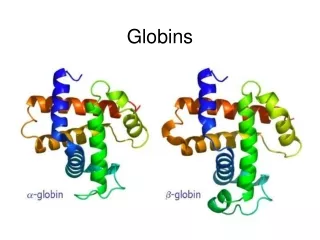
Globins
Globins. Lecture 10/01/2009. The Backbone structure of Myoglobin. 2. Myoglobin: 44 x 44 x 25 Å single subunit 153 amino acid residues 121 residues are in an a helix. Helices are named A, B, C, …F. The heme pocket is surrounded by E and F but not B, C, G, also H is near the heme.


Globins
E N D
Presentation Transcript
Globins Lecture 10/01/2009
The Backbone structure of Myoglobin 2 Myoglobin: 44 x 44 x 25 Å single subunit 153 amino acid residues 121 residues are in an a helix. Helices are named A, B, C, …F. The heme pocket is surrounded by E and F but not B, C, G, also H is near the heme. Amino acids are identified by the helix and position in the helix or by the absolute numbering of the residue.
Helix E Distal His Fe3+ - O O - Fe3+ Proximal His Helix F • Role of the Globin • Modulate oxygen binding affinity • Make reversible oxygen binding possible By introducing steric hindrance on one side of the heme plane interaction can be prevented and oxygen binding can occur. A heme dimer is formed which leads to the formation of Fe(III)
The Heme group Each subunit of hemoglobin or myoglobin contains a heme. - Binds one molecule of oxygen - Heterocyclic porphyrin derivative - Specifically protoporphyrin IX The heme prosthetic group in Mb ad Hb: protoporphyrin IX + Fe(II) The iron must be in the Fe(II)form or reduced form (ferrous oxidation) state.
E7 F8
Hemoglobin Spherical 64 x 55 x 50 Å two fold rotation of symmetry a and b subunits are similar and are placed on the vertices of a tetrahedron. There is no D helix in the a chain of hemoglobin. Extensive interactions between unlike subunits a2-b2 or a1-b1 interface has 35 residues while a1-b2 and a2-b1 have 19 residue contact. Oxygenation causes a considerable structural conformational change
Oxygenation rotates the a1b1 dimer in relation to a2b2 dimer about 15° The conformation of the deoxy state is called the T state The conformation of the oxy state is called the R state individual subunits have at or r if in the deoxy or oxy state. What causes the differences in the conformation states?
The positive cooperativity of O2 binding to Hb The effect of the ligand-binding state of one heme on the ligand-binding affinity of another. The Fe iron is about 0.6 Å out of the heme plane in the deoxy state. When oxygen binds it pulls the iron back into the heme plane. Since the proximal His F8 is attached to the Fe this pulls the complete F helix like a lever on a fulcrum.
Hemoglobin structure DeoxyHb • b-monomers are related by 2-fold symmetry (same is true for a) • Note changes in structure: • between b-monomers – see big double-headed arrows • at points of contact – see small arrows Binding of the O2 on one heme is more difficult but its binding causes a shift in the a1-b2 (& a2-b1) contacts and moves the distal His E7 and Val E11 out of the oxygen’s path to the Fe on the other subunit. This process increases the affinity of the heme toward oxygen. The a1-b2 contacts have two stable positions . These contacts, which are joined by different but equivalent sets of hydrogen-bonds that act as a binary switch between the T (deoxy) and the R (oxy) states oxyHb
The major structural difference between the quaternary conformations of (a) deoxyHb and (b) oxyHb Note: This view is from the right side relative to the previous slide.
The positive cooperativity of O2 binding to Hb The effect of the ligand-binding state of one heme on the ligand-binding affinity of another. The Fe iron is about 0.6 Å out of the heme plane in the deoxy state. When oxygen binds it pulls the iron back into the heme plane. Since the proximal His F8 is attached to the Fe this pulls the complete F helix like a lever on a fulcrum.
Mechanism of Cooperativity in Hemoglobin • T-state (deoxyhemoglobin) • Fe is 0.6 Å out of heme plane • R-state (oxyhemoglobin) • Fe is in the heme plane • Helix containing F8 shifts • Change in quaternary structure • C-terminal residues (Arg141a, His146b, and Val1a) change interactions and/or ionization state (Bohr effect)
Binding causes a shift in the a1-b2 contacts and moves the distal His E7 and Val E11 out of the oxygen’s path to the Fe on the other subunit. This process increases the affinity of the heme toward oxygen. The a1-b2 contacts have two stable positions with different but equivalent sets of hydrogen bonds to act as binary switch between the T and the R states
a. Free energy changes with fractional saturation b. Sigmoidal binding curve as a composite of the R state binding and the T state binding.
Association kinetics of O2 binding to myoglobin Written backwards we can get the dissociation constant Fractional Saturationsolve for [MbO2] and plug in
The Hill Equation E = enzyme, S = ligand, n= small number This is for binding of 1 or more ligands O2 is considered a ligand 2. 1. Fractional Saturation = bound/total
Hill Plot Rearrange equation 4. y = mx + b n = slope and x intercept of -b/m
The visible absorption spectra for hemoglobin The red color arises from the differences between the energy levels of the d orbitals around the ferrous atom. Fe(II) = d6 electron configuration low spin state Binding of oxygen rearranges the electronic distribution and alters the d orbital energy. This causes a difference in the absorption spectra. Bluish for deoxy Hb Redish for Oxy Hb Measuring the absorption at 578 nm allows an easy method to determine the percent of O2 bound to Hb
Things to remember Hb subunits independently compete for O2 for the first oxygen molecule to bind When the YO2 is close to 1 i.e. 3 subunits are occupied by O2 , O2 binding to the last site is independent of the other sites However by extrapolating slopes: the 4th O2 binds to hemoglobin 100 fold greater than the first O2 A DDG of 11.4 kJ•mol -1in the binding affinity for oxygen When one molecule binds, the rest bind and when one is released, the rest are released.
Contrast Mb O2 binding to Hemoglobin YO2 = 0.95 at 100 torr but 0.55 at 30 torr a DYO2 of 0.40 Understand Fig 9-3 Hb gives up O2 easier than Mb and the binding is Cooperative!!
Allosteric Proteins Symmetry model (Monod-Wyman-Changeux) Chapter 12 Enzymes!! Sequential model (Koshland)
The Bohr Effect Higher pH i.e. lower [H+] promotes tighter binding of oxygen to hemoglobin and Lower pH i.e. higher [H+] permits the easier release of oxygen from hemoglobin Where n = 0, 1, 2, 3 and x 0.6 A shift in the equilibrium will influence the amount of oxygen binding. Bohr protons
CO2 Transport and The Bohr Effect Higher pH i.e. lower [H+] (more basic) promotes tighter binding of oxygen to hemoglobin and Lower pH i.e. higher [H+] (more acidic) permits the easier release of oxygen from hemoglobin Where n = 0, 1, 2, 3 and x 0.6 A shift in the equilibrium will influence the amount of oxygen binding. Bohr protons
Origin of the Bohr Effect The T R transition causes the changes in the pK’s of several groups. The N-terminal amino groups are responsible for 20-30% of the Bohr effect. His146b accounts for about 40% of the Bohr effect salt bridged with Asp 94b. This interaction is lost in the R state. Networks of H-bonds & ion pairs in T-state • The T-state is shown above. • TR transition causes breakage of terminal interactions and changes in ionization states of His146b and Val1a (part of Bohr effect)
Look at the relation between pH and the p50 values for oxygen binding. As the pH increases the p50 value decreases, indicating the oxygen binding increases (opposite effect,when the pH decreases). At 20 torr 10% more oxygen is released when the pH drops from 7.4 to 7.2!! The Bohr effect: Importance in transporting O2 and CO2 As oxygen is consumed CO2 is released. Carbonic Anhydrase catalyzes this reaction in red blood cells. • 0.6H+ released for each O2 binding • CO2 + H2O H+ + HCO3-, catalyzed by carbonic anhydrase – main mode of elimination of CO2
D-2,3-bisphosphoglycerate (BPG) BPG binds to Hb (deoxy state) and decreases the O2 affinity and keeps it in the deoxy form. BPG binds 1:1 with a K=1x10⁻⁵ M to the deoxy form but weakly to the oxy form Fetal Hb (a2g2) has low BPG affinity b-His143 to Ser in g chain
The P50 value of stripped hemoglobin increases from 12 to 22 torr by 4.7 mM BPG
At 100 torr or arterial blood, hemoglobin is 95% saturated At 30 torr or venous blood, hemoglobin is 55% saturated Hemoglobin releases 40% of its oxygen. In the absence of BPG, little oxygen is released. Between BPG, CO2, H+, and Cl- all O2 binding is accounted for.
BPG levels are partially responsible for High-Altitude adaptation BPG restores the 37% release of O2 at higher elevations between arterial and venous blood
Fetal Hemoglobin • Fetal hemoglobin has a different b subunit called a g subunit or a2g2. • InFetal hemoglobin, BPG does not affect this variant and the baby’s blood will get its oxygen from the mothers hemoglobin. • The transfer of oxygen is from the mother (less tightly bond) to the baby (more tightly bond).
Sickle Cell Mutation • Glu 6 ---> Val 6 mutation on the hemoglobin b chain • Decreases surface charge • More hydrophobic • Frequency 10% USA versus 25% in africa. • Forms linear polymers
Normal and sickled erythrocytes Heterozygotes carrying only one copy of the sickle-cell gene are more resistant to malaria than those homozygous for for the normal gene.
Hemoglobin mutants • There are about 500 variants of hemoglobin 95% are single amino acid substitutions. • 5% of the world’s population carries a different sequence from the normal. • Changes in surface charge • Changes in internally located residues • Changes stabilizing Methemoglobin (oxidized Fe(III)) • Changes in the a1-b2 contact • Changes in surface rarely change the function of hemoglobin with the exception of the sickle cell mutation. • Internal residues cause the hemoglobin to contort to different shapes and alter its binding properties. Heinz bodies are precipitated aggregates of hemoglobin. Usually cause hemolytic anemia characteristic by cell lysis.