Muscle Structure and Function
This guide delves into the complexities of muscle biology, focusing on the structure and function of skeletal, smooth, and cardiac muscles. It covers essential topics such as major muscle groups, histology, gross and microscopic anatomy, the sliding filament model of contraction, and the biochemical processes underlying muscle contraction. Additionally, it explains the role of neuromuscular junctions, action potentials, and energy production pathways, including aerobic and anaerobic respiration, crucial for muscle metabolism.

Muscle Structure and Function
E N D
Presentation Transcript
Muscle Structure and Function Biology 2121 Chapters 9-10
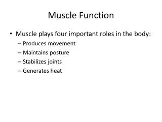
Introduction 1. Functions • Movement, Posture; Heat and Joints 2. Naming (321) • Location; Shape (temporalis; deltoid) • Size (gluteus maximus) • Fiber Direction (abdominus rectus) • Origins (bicep) • Location of Attachment (sternocleidomastoid)
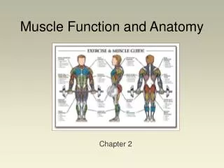
Major Muscle Groups Chest and Shoulder Group Abdominals Quadriceps and Hamstring Group
Histology of Muscle Tissue- Skeletal Muscle • Voluntary • Found in major muscle groups • Striated • Multinucleated • Peripheral
Smooth Muscle • Involuntary • Found in digestive system organs, bladder, etc. • Non-Striated • Uninucleated • Spindle-Shaped nuclei
Cardiac Muscle • Involuntary • Heart only • Branching Fibers • Uni-Nucleated
Gross Anatomy • 1. Wholemuscle • Epimysium • 2. Fascicles • Perimysium • 3. MuscleFiberCells • Long and multi-nucleated • Sarcolemma and sarcoplasma • Endomysium
Attachments • Tendons • Aponeurosis • Direct or Indirect Attachments
Microscopic Anatomy – Myofibrils • 1. Contractileproteins • 2. Sarcomeres • Actin and myosin • 3. Myofilaments • Actin(thin) • Myosin (thick)
Sarcomere • 1. A ‘sarcomere’ • Z to Z • I-Band (light zone) • A-Band (dark zone) • 2. StructuralProteins • Elastic filaments – Titan • 3. SlidingFilamentModelof Contraction • Link
Chemical Stimulation and Muscle Contractions • 1. Stimulation and Neurotransmitters • Acetylcholine • 2. NeuromuscularJunction • Junction – muscle/nerve interface • Separation – “synapse”
Events at the Neuromuscular Junction • 1. Nerve Impulse • 2. Calcium ions – Axon Terminal of Nerve • 3. Vesicle and release of ACh • 4. ACh receptors and Acetylcholinesterase • 5. Sodium-Potassium exchange • 6. Action Potential formed
Action Potential • 1. RestingMembrane • -70 mV • Na+ and K+ • 2. Reversalof Charges • Depolarization • 3. Movesin oneDirection
Excitation and Contraction • 1. ActionPotential moves along the sarcolemma • 2. Down the T-Tubule • 3. SarcoplasmicReticulum and Release of Ca++ • 4. Ca++ moves to the sarcomere
Formation of a Cross-Bridge • 1. Ca++ interacts with troponin • 2. Removestropomyosin • 3. Allows for MyosinHeadAttachment • 4. Formation of a cross-bridge
Cross-Bridge Cycling • 1. Myosinheadsattach forming cross- bridge • 2. WorkingStroke • 3. ATPbreaks cross- bridge • 4. ATPhydrolysis • 5. High-energy configuration – New Cross-bridge
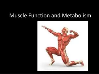
ATP and Muscle Metabolism ATP Functions – Driving Cellular Work
ATP Production 1. CreatinePhosphate – Quick and Fast!!! 2. AnaerobicRespiration • “Lactic Acid Fermentation” • 1 glucose molecule = 2 ATP Net 3. AerobicRespiration • 1 glucose molecule = 36 ATP Net • Mitochondria of the Cell
Fermentation • Glycolysis • (2) Pyruvicacidmolecules (3-C) • Bloodflowrestrictedduring vigorous exercise (low oxygen) • Lacticacidformed • Anaerobic glycolysis