Download
1 / 21
220 likes | 470 Vues
Pelvis & Thigh Injuries. Chapter 8 p.272. Clinical Anatomy p. 272. Bony Anatomy Ileum, ischium, pubis ASIS/PSIS Labrum Sacroiliac joints Bursae (p.279) Trochanteric Ischial. History – p.279. Location of symptoms Table 8-2, p. 281) “hip pain” Hip, SI, lumbar, or referred pain

E N D
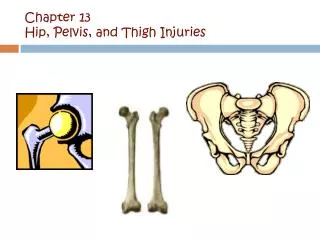
Pelvis & Thigh Injuries Chapter 8 p.272
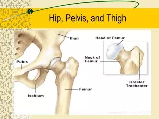
Clinical Anatomyp. 272 • Bony Anatomy • Ileum, ischium, pubis • ASIS/PSIS • Labrum • Sacroiliac joints • Bursae (p.279) • Trochanteric • Ischial
History –p.279 • Location of symptoms • Table 8-2, p. 281) • “hip pain” • Hip, SI, lumbar, or referred pain • Anterior pain • Usu. Muscle strain of adductors/flexors • Pubic pain • Referred pain or adductor strain
History –p.281 • Onset: • usually gradual/chronic • Correlates with change in activity • Mechanisms: • Sudden ECC contraction (avulsion fx) • Sudden trauma to PSIS (hip pointer) • Trauma to buttocks (contusion or fx) • PMH: • Congenital defectsaltered biomechanics • Prior injuriesstiffness
Inspection/Observationp. 280 • Inspection difficult • Observe biomechanics • Q-angle • 13º (m) • 18 º (f) • 8º in 90º knee flex. • Angle of torsion • WNL=8º -15º • Retroversion (ER) • Anteversion (IR) • Box 8-1, p. 282
Observation/Inspection • Iliac crest • hip pointer • Fig. 8-13, p. 282 • Leg length (p. 209) • True leg length: • ASIS to MM • Apparent leg-length • Umbilicus to MM • If TLL is (-) and ALL is (+) • suspect pelvic obliquity or adductor contracture
Observation/Inspection:Angle of Inclination—p.281 • NL= 125° • >125 °= coxa valga • Genu varum or lateral patellae • <125 °=coxa vara • Genu valgum or • Medial patellae • Confirm with x-ray
Palpationp. 283 • Iliac crest • ASIS/PSIS • Greater trochanter • Rectus femoris • SI joints • Hamstrings
Functional Testingp. 285 • ROM: • Flexion: • 120º- 130º • Extension • 10º-30º • Abduction • ~45º • Adduction • 20º - 30º • IR • 35º - 45º • ER • 40º - 50º
SI Joint Functional Testing • Normal motion: • Sitting • PSIS moves sup/ant in lumbar ext • PSIS moves inf/post in lumbar flex • Lateral side-bending • PSIS should elevate and move medially
Pathologiesp. 294 • Muscle strains • Quadriceps contusion • Bursitis • Piriformis syndrome • Sacroiliac dysfunction
Muscle Strainsp. 294 • Quadriceps/ Hamstrings/ Adductors/ Hip flexors • Mechanisms: • High ECC loading • Overuse/ Overtraining • Strength imbalance • Isolate the injury • Pain increases with AROM or PROM • (-) joint pain/laxity • Table 8-4, p. 294
Quadriceps Contusionp. 299 • Trauma to ant. Quadriceps (“Charley Horse”) • Inc. risk of Myositis Ossificans • Painful to touch with ecchymosis and edema • Atrophies quickly • Tx: • Ice/stretch • Strengthen • Protect from re-injury
Bursitisp. 295 • Caused by inc. friction between 2 tissues • “Snapping Hip” syndrome • Mechanisms: • Direct trauma • Biomechanical/ congenital factors • Prolonged sitting • Sites: • Trochanteric • Ischial
Piriformis syndromep. 298(Table 8-7) • Sciatic nerve impingement at piriformis muscle • Entrapped by spasm/ inflammation/ hypertrophy/trauma • Mimics lumbar nerve root injury • Tenderness @ sciatic notch • Antalgic gait • Table 8-7, p. 298 • Numbness/ Paresthesia which inc. with activity • Piriformis tenderness • More common in runners & females • Dx. Frequency decreasing
Osteitis Pubis • Caused by repeated overload to adductors, usually from running activities • Often confused with ‘groin strain’ • S/S – gradual onset, aggravated with running, kicking, pivoting on one leg • Increased pain with abdominal exercises • Tx – NSAIDS, rest, LIGHT stretching, hydrotherapy, alternative aerobic activity, adductor strengthening • May require 2-3 months to recover
Special Tests for the Hip/Pelvis • Thomas test • Trendelenburg’s test • Hip Scouring test
Thomas Testp. 289 • Positive (+)Test= • tight hip flexors OR • Tight rectus femoris • Positioning: • Supine, with knees flexed over end of table • Procedure: • Flex one hip to chest and watch opposite leg • Contralateral extremity should not be affected
Trendelenburg's Testp. 293, Box 8-5 • Tests for hip weakness • Muscular (g. medius) • Neurologic • Position: • Stand on one leg • Procedure • Look for shift in BW to maintain balance • (+)=Pelvis lowers on non-weight-bearing side
Hip Scouring Testp. 297,(Box 8-6) • Tests integrity of articular cartilage of hip • Similar to McMurray test for the knee • Position: • Supine with knee & hip flexed fully • Procedure: • IR/ER femur in multiple hip flexion angles • (+)= reproduced pain in hip with crepitus