Download
1 / 22
220 likes | 237 Vues
Practice identifying brain structures, sinuses, glands, muscles, and more in MRI images. Learn key anatomical features and diagnostic clues.

E N D
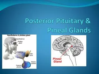
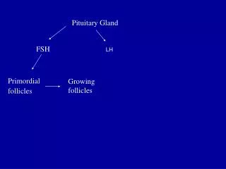
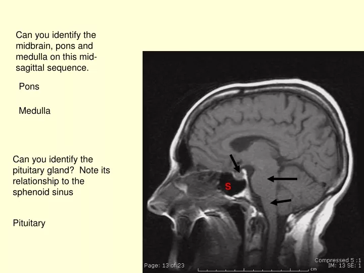
Can you identify the midbrain, pons and medulla on this mid-sagittal sequence. Pons Medulla Can you identify the pituitary gland? Note its relationship to the sphenoid sinus S Pituitary
Can you identify the corpus callosum and the cerebellum? Corpus callosum cerebellum
This is a coronal T1 weighted sequence through the brain. Can you identify the lateral ventricles? What are these glandular structures noted laterally (red arrows)? Parotid glands
What is this bony structure (red arrow)? See the adjacent muscular attachments. Mandible What are these circular low signal structures (yellow arrows). Hint flowing blood can look like low signal, sometimes called “flow voids”. Cavernous carotids
What is this large are of signal void, in the middle of the image (arrows)? Hint, air produces no signal. Sphenoid sinus
What is this muscular structure (yellow arrows)? This is the tongue. Do you see the areas of high signal in the muscle. We always see fat (high signal on T1) in the tongue
Can you identify the maxillary sinuses? Hint, air filled sinuses will produce no signal Can you identify the maxillary sinuses? Hint, air filled sinuses will produce no signal Can you identify the nasal turbinates?
Now let’s take a look at the orbits. Try and pick out the optic nerve, superior rectus, superior oblique, medial, lateral and inferior rectus extra-ocular muscles. The inferior oblique muscle is note seen on these images. Blue=optic nerve, White=inferior rectus Red=medial rectus, Yellow=superior oblique Green=superior rectus, Pink=lateral rectus Review the innervation of the these muscles. Which 2 muscles are not innervated by the oculomotor nerve (III) Superior oblique– trochlear nerve (IV) Lateral rectus– Abducens nerve (VI)
What is the signal void over the orbits (arrow)? Frontal sinuses
These are coronal contrast enhanced images, do you see the carotid arteries in the cavernous sinuses. Note the venous blood in the sinuses is slow flowing and in bight on these images but the blood in the carotid arteries is fast flowing and it shows up as signal void. Cavernous sinuses Carotids
Now we are out of the cavernous sinus. Do you see the abnormal enhancing lesion in this case?
What are the muscles attached to the inside and the outside of the mandible? Can you identify the mandible? Masseter muscle Medial pterygoid muscle
Do you see the right parotid gland? Why don’t you see the left parotid gland? (was he born without one) Unlikely, his head is just tiled in the MRI scanner so the left one is out of the plane
Can you identify the air filled spaces (areas of signal void) on this image? Mastoid air cells Nasopharynx Maxillary Sinuses
What is this blood filled normal venous structure? Transverse sinuses
What extra-ocular muscles do you see on this image? Do you see any areas of abnormal enhancement? Inferior rectus on the right (yellow arrow) Lateral rectus on the left (red arrow) This is the lesion, adjacent to the cavernous extending to the orbital apex
What extra-ocular muscles can you identify on the right? Do you see the optic nerve? Medial rectus blue arrow Lateral rectus red arrow Optic nerve pink arrow
Can you identify the frontal sinuses and the sylvian fissures. Frontal sinuses pink arrows Sylvian fissures blue arrows