Download
1 / 85
1.01k likes | 1.51k Vues
Trans-esophageal & Intra-cardiac Echocardiography. Himal raj Sr cardiology. HISTORY. Side and Gosling ( 1971) - TEE for CwD of cardiac flow Frazin et al (1976 ) - TEE M mode echo Hisanaga et al ( 1977) - illustrated use of cross sectional real time imaging. introduction.

E N D
Trans-esophageal & Intra-cardiac Echocardiography Himal raj Sr cardiology
HISTORY • Side and Gosling (1971) -TEE for CwD of cardiac flow • Frazin et al(1976) -TEE M mode echo • Hisanaga et al (1977) -illustrated use of cross sectional real time imaging
introduction • TEE uses sound waves to create high-quality moving pictures of heart and its blood vessels • involves a flexible tube or probe with a transducer at its tip • probe is guided down throat and esophagus • more detailed pictures of heart as esophagusis directly behind heart
TEE: Types • Types of TEE : • 2-Dimensional (2D) • 3-Dimensional (3D) • Standard TEE pictures are 2D • 3D pictures provide more details about • Structure and function of heart and Its blood vessels • 3D TEE helps to diagnose heart problems like: • Congenital heart disease • Heart valve disease and • To assist with heart surgery
TEE: Advantages • Transducer - 2- 3 mm from heart • Closer to posterior structures- Better visualization of LA, LAA, PV, MV, LV, Aorta • Far from surgical area - Intra-operative monitoring • High resolution images : [Absence of intervening lung or bone tissue - Better signal to noise ratio and decreased image depth – allows use of higher freq (5 and 7 MHz) transducers – enhances image quality]
TEE: DISADVANTAGES • semi invasive procedure: chances of injury ; • needs special setup, technique, preparation, instrumentation • needs orientation and expertise
Indications -common • Assessment of prosthetic valves; infective endocarditis ; native valve disease • Assessment of a suspected cardioembolicevent • Assessment of cardiac tumors • Assessment of atrial septal abnormalities • Assessment of aortic dissection, intramural hematomas • Evaluation of CHD; CAD ;pericardial disease • Evaluation of critically ill patients • Intraoperative monitoring • Monitoring during interventional procedures • Stress echocardiography • Nondiagnostic TTE
Contraindications ABSOLUTE • Oesophageal stricture or obstruction • Suspected or known perforated viscus • Instability of cervical vertebrae • GI bleeding not evaluated RELATIVE • Esophageal varices or diverticula • Cervical arthritis • Oropharyngeal distortion • Bleeding diathesis or over-anticoagulation
procedure • 4- 6 hours fasting • Written consent • Intravenous line ; oxygen ; suction equipment ; Remove denture or devices ; 2% lidocaine spray • ECG must be monitored throughout • Left lateral position • Introduce the probe with some anteflexion through a bite block
procedure • Routine antibiotic prophylaxis before TEE is not advocated [ risk of IE is extremely low].Recommended in high risk patients - prosthetic valves, multivalvularinvolvement or those with a past h/o IE] • Persistent resistance to advancing the instrument mandates termination of TEE and endoscopy should be performed before re-examination. • After each TEE - Disnfect ; Check for any damage - ensure electrical safety
complications • Majority are minor. • Major complications [death, laryngospasm, sustained VT & CHF occur in ≈ 0.3% of patients] • Cardiac complications include SVT or AF, VT, bradycardia, transient hypotension or hypertension, angina ,CHF and pulmonary edema.
complications MAJOR • Death • Esophageal rupture • Laryngospasm or bronchospasm • Congestive heart failure or pulmonary edema • Sustained ventricular tachycardia
complications MINOR • Excessive retching or vomiting • Sore throat • Hoarseness • Minor pharyngeal bleeding • Blood tinged sputum • Non sustained or sustained supraventricular tachycardia • Atrial fibrillation • Nonsustained ventricular tachycardia • Bradycardia or heart block • Transient hypotension • Transient hypertension • Angina • Transient hypoxia • Parotid swelling • Tracheal intubation
TEE PROBE • Modification of standard gastroscope, with transducers in place of fibreoptics • Conventional rotary controls with inner and outer dials • Inner dial guides anteflexion and retroflexion • Outer dial controls medial and lateral movement • Multiplane probe has a lever control to guide rotation
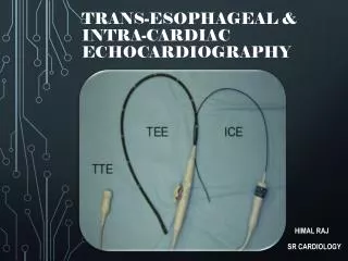
TEE Transducer TEE Transducer Relation of TEE transducer with heart
TEE PROBE • Monoplane TEE - provides images in horizontal plane only • Biplane TEE - orthogonal longitudinal plane also • Multiplane TEE transducer : • single array of crystals [phased array transducers with 64 -256 piezoelectric elements] • that can be electronically and mechanically rotated in an arc of 180 ° • to produce a continuum of transverse and longitudinal images from a single probe position
Standard imaging plane levels (from the incisors) • upper or high esophageal (25–28 cm) • mid-esophageal (29–33 cm) • transgastric(38–42 cm) • deep-transgastric(>42 cm)
PROCEDURE Proceed systematically - from mid esophagus [≈35 cms from the incisors] to gradually more distal esophagus, fundus of the stomach after gentle advancement across the cardia [≈40-50 cms from incisors] and finally slow withdrawal of the probe for complete scan of the thoracic aorta [from high esophageal views].
PROCEDURE • A complete TEE exam usually takes 15–20 min. • An abbreviated or problem-focused TEE study may be appropriate in unstable or uncooperative patients
Transducer manipulation options • [1] Advancement/withdrawal(for inferior or superior structures respectively) • [2] Rotation(clockwise to view rightward structures and counter- clockwise for leftward structures)
Transducer manipulation options • [3] Anteflexion and retroflexion of the probe shaft (to view structures towards the heart base or towards the apex) • [4] Leftward and rightward flexion of the probe shaft(used infrequently with the advent of multiplane probes)
Transducer manipulation options • [5] Electronic image plane rotation(0–1800)
By convention, in TEE, tip of 2D sector is displayed on top of screen and left-sided cardiac structures appear on right side of display.
Prior guidelines developed by the ASE and the SCA have described the technical skills for acquiring 20 views in the performance of a comprehensive intraoperative multiplanetransesophagealechocardiographic examination • But current guidelines recommend that a basic PTE examination should focus on encompassing the 11 most relevant views.
Cross-sectional views of the 11 views of the ASE and SCA basic PTE examination.
Mid Esophagus 4C ( 0°) Position probe in mid-esophagus behind LA. depth 14cm, angle 0-10°. Image all 4 heart chambers. Optimize LV apex by slight retroflexion of probe tip. Ensure no part of AV or LVOT is seen. Aim to maximize TV diameter, and adjust depth to view entire LV. Assess :chamber size; ventricular function; mitral valve disease; tricuspid valve disease; ASD; pericardial effusion
ME 2C ( 90° ) From ME 4C : keep probe tip still and MV in the center; rotate omniplane angle forward to 80-100°; RA + RV disappear, LAA appears.Retroflex probe tip for true LV apex; adjust depth to see entire LV apex. Assess : LAA mass/thrombus; LV size and function; MV disease (A1, A2 & P3 scallops); MV annulus measurement ‘
ME LAX (120°) Rotate omniplane angle forward to 120-130° Imaging plane is directed thru the LA to image the aortic root in LAX and entire LV. The more cephalad structures are lined up on the display right. The LV anteroseptal + inferolateral walls & MV segments, A2 and P2 are seen. Assess : LV function, MV disease, AV and aortic root disease, IVS pathology.
ME Asc A LAX ( 90°) Find the ME AV LAX (120°). Withdraw the probe to bring the right pulmonary artery in view Decrease omniplane angle slightly by 10-20° to make the aortic wall symmetric Imaging plane is directed thru the right pulmonary artery to image the proximal ascending aorta in LAX. For: aortic pathology, pericardial effusion, pulmonary embolus
ME Asc A SAX (0°) From ME AV LAX (120°) OR from ME AV SAX (30°)…. Withdraw probe (asc aorta ), Rotate the omniplane angle back to 0° Imaging plane is directed slightly above the aortic valve thru the RPA(seen in LAX), ascending aorta (seen in SAX) and SVC (SAX). For : PA pathology, pulmonary embolus, ascending aorta pathology ,PDA, swan-ganz in SVC
ME AV SAX (30-45°) From ME 4C (0°) withdraw cephalad to obtain the ME 5C(0°) [imaging plane is directed thru the LA and aligned parallel to the AV annulus] rotate to 30-45°;center aortic valve and aim to make 3 aortic valve cusps symmetric. Withdraw probe for coronary ostia.Advance probe for LVOT. Assess : AV disease, OS ASD, LA size, coronary artery pathology
ME RVIO View (60-75°) From ME AV SAX (30-60°) rotate omniplane angle to 60- 75° Optimize TV leaflets, open up RVOT, Bring PV + main PA into view For : P valve / PA / RVOT /TV pathology /VSD
ME BCV ( 90°) From ME 2 C (90°), Turn entire probe right Change angle or rotate probe slightly to image both IVC (left) and SVC (right) simultaneously For : ASD (secundum, sinus venosus), atrial pathology, lines/wires,Venous cannula (SVC, IVC)
TG mid SAX (0°) Advance probe until you see stomach (rugae) or liver. anteflex to contact stomach wall and inferior wall of heart . center LV by turning probe R or L . image both papillary muscles . imaging plane transversely thru the mid inferior wall of the LV with all 6 mid LV segments viewed at once from the stomach. For: Left ventricle size, function, IVS motion, VSD, pericardial effusion