Acute Pancreatitis
Acute Pancreatitis. Dr. Belal M. Hijji, PhD, RN April 2 & 4, 2011. Learning outcomes. By the end of this lecture, students will: Have an introductory statement about acute pancreatitis. Identify clinical manifestations. Describe diagnostic evaluation.

Acute Pancreatitis
E N D
Presentation Transcript
Acute Pancreatitis Dr. Belal M. Hijji, PhD, RN April 2 & 4, 2011
Learning outcomes • By the end of this lecture, students will: • Have an introductory statement about acute pancreatitis. • Identify clinical manifestations. • Describe diagnostic evaluation. • Discuss medical and nursing management.
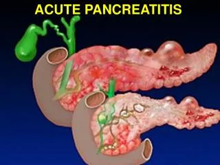
Introduction to Acute Pancreatitis • Acute pancreatitis ranges from mild, self-limited disorder to a sever, rapidly fatal disease that does not respond to any treatment. • Mild acute pancreatitis is characterised by edema and inflammation confined to the pancreas. The patient is acutely ill, at risk for hypovolemic shock, fluid and electrolyte imbalance, and sepsis. • Severe acute pancreatitis is characterised by complete enzymatic digestion of the gland. Enzymes damage local blood vessels and bleeding and thrombosis can occur. In this condition, local complications (i.e. pancreatic cysts, abscesses, acute fluid collection) and systemic complications (i.e. hypoxia, shock, renal failure, GI bleeding) can occur.
Clinical Manifestations • Severe abdominal pain causes patient to seek medical care. Pain is midepigastric or periumbilical; it occurs 24-48 hours after a heavy meal or alcohol ingestion, and it may be diffuse and difficult to localise. • The patient looks ill with abdominal guarding. • Purplish discoloration of the flank or around the umbilicus may indicate severe pancreatitis. • Decreased peristalsis, nausea and vomiting. • Fever, jaundice, and mental confusion. • Hypotension reflects hypovolemia and shock caused by loss of large amounts of protein-rich fluids into the tissues and peritoneal cavity. • Respiratory distress, hypoxia, tachypnea, and abnormal blood gas values.
Diagnostic Evaluation • History of abdominal pain. • Presence of risk factors. • Physical examination (abdominal distention, abdominal mass, decreased peristalsis) • Diagnostic tests: • Elevated serum amylase and lipase. • Elevated urinary amylase. • Leukocytosis. • Transient hyperglycemai & glucosuria. • High serum bilirubin. • Ultrasound and CT scan of abdomen identify an increase in the diameter of the pancreas, and to detect cysts and abscesses. • Hematocrit and hemoglobin.
Medical Management • Patient is kept on nothing per os (NPO) to inhibit the stimulation of pancreas and its secretion of enzymes. • ُEnteral or parenteral nutritional support (see next 2 slides). • Histamine – 2 (H2( antagonists such as cimetidine (tagamet) or ranitidine (Zantac) to limit pancreatic activity by inhibiting gastric acid secretion. • Pain management by using opoids (Morphine). • Intensive care to correct fluid, blood loss, and low albumin. This is important for maintaining fluid volume and preventing renal failure. • Respiratory care to correct hypoxemia. This care may range from monitoring of ABG to use of humidified oxygen to intubation and mechanical ventilation.
Subclavian triple-lumen catheter used for paenteral nutrition and other therapies. (A) The catheter is threaded through the subclavian vein into the vena cava. (B) Each lumen is an avenue for solution administration
Surgical intervention to assist in the diagnosis of pancreatitis, to establish pancreatic drainage, and to debride a necrotic tissue. Multiple sump tubes are used after pancreatic surgery. Triple-lumen tubes consist of ports that provide tubing for irrigation, air venting, and drainage.
Nursing Management • Pain relief and patient comfort • Administration of prescribed pain killers (parenteral morphine). • Assessment of pain (slides 12 & 13) and effectiveness of the pharmacologic and nonpharmacologic interventions (proper positioning, music, distraction, imagery). • Keeping patient NPO • Frequent oral care to relieve discomfort and dryness of mouth. • Bed rest to decrease the secretion of gastric and pancreatic enzymes. • Reporting increased level of pain to physician (This may indicate hemorrhage or inadequate analgesia).
Improving breathing pattern • Place the patient in semi-Fowler’s position (slide 8). • Frequent position changes to prevent atelectasis (lung collapse) and pooling respiratory secretions. • Frequent deep breathing, coughing exercises, and incentive spirometry to improve respiratory function.
Improving nutritional status • Assessment of patient nutritional status (History of weight loss, body mass index (BMI) (next slide)). • Laboratory tests (albumin, transferrin, prealbumin) and daily weight. Albumin & transferrin are decreased in malnutrition, while prealbumin is elevated. • Administration of enteral or parenteral nutrition. • Monitoring blood glucose level every 4-6 hours. • When oral feeding is allowed, the nurse provides high carbohydrate low protein low fat diet. • Educating the patient about the importance of avoiding heavy meals and alcohol.
Weight in Kilos BMI for Men Height in Metres normal weight (bmi 20-25) overweight (bmi>25) underweight (bmi<20)
Maintaining skin integrity • The patient is at risk of developing pressure ulcer because of nutritional deficiencies, bed rest, and restlessness. • The nurse assesses the wound, drainage sites, and skin for signs of infection, inflammation, and break down. • The nurse carries out wound care as prescribed, and protects intact skin from contact with drainage. • The nurse performs position changes every 2 hours.
Monitoring and managing potential complications • For fluid and electrolytes disturbances, the nurse should assess the patient’s fluid and electrolytes status by noting skin turgor and moistness of mucus membranes. Daily weight, recording intake and output, are daily abdominal girth if ascites is present, are another nursing interventions. • The nurse administers intravenous fluids possibly with the infusion of blood or blood products. The nurse promptly reports low BP and reduced urine output, which indicate hypovolemia and shock or renal failure. • The nurse assesses vital signs