Acute Pancreatitis
230 likes | 1.01k Vues
Acute Pancreatitis. Ashley Duckett, MD Theresa Cuoco, MD, FACP. Objectives. Identify clinical presentation and etiologies of acute pancreatitis Recognize the importance of severity of pancreatitis in determining management and outcomes

Acute Pancreatitis
E N D
Presentation Transcript
Acute Pancreatitis Ashley Duckett, MD Theresa Cuoco, MD, FACP
Objectives • Identify clinical presentation and etiologies of acute pancreatitis • Recognize the importance of severity of pancreatitis in determining management and outcomes • Understand the indications for imaging and antibiotics in acute pancreatitis
Key Messages • Early aggressive hydration is critical in the management of acute pancreatitis. • Imaging with contrasted CT or MRI is indicated at 48-72 hours in patients with severe disease. • Early enteral nutrition improves outcomes in patients with severe pancreatitis. • Severity scores can be used to triage patients at risk for complications.
Clinical presentation • Abdominal pain, distention • Nausea/vomiting (90%) • Fever, tachycardia, shock, coma • Dyspnea (pleural effusion) • Shallow respirations (diaphragmatic irritation) • Jaundice • Grey-Turner’s/Cullen’s 1% - intraabdominal hemorrhage
Etiologies • Gallstones- 35-40% (increased ALT >150 is 50% sensitive and 96% specific) *women • Alcohol – 30% *men • Hypertriglyceridemia • Hypercalcemia • Post-ERCP -3% of diagnostic, 25% if SOD studies • Drugs – direct toxic, immunologic, ischemic causes – cocaine, HCTZ • Genetic mutations • Trauma • Infection – Viruses (mumps, hep B, CMV, HIV), Bacteria (mycoplasma, legionella, salmonella), fungi, parasites • Idiopathic -15-20% • Autoimmune (although usually presents mimicking neoplasm) • Scorpion bite • SOD dysfunction? Pancreatic divisum?
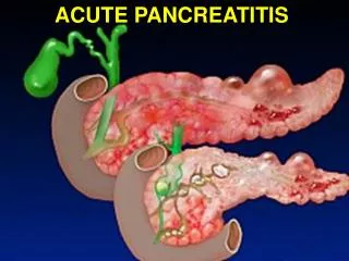
Pathophysiology • Activation of trypsin in acinar cells • Activating multiple pancreatic digestive enzymes (synthesized but not secreted) • Leak into interstitial space and systemic circulation • Intrapancreatic Inflammation • Mediated by cytokines/inflammatory markers • SIRS • Extrapancreatic Inflammation • ARDS • Multiple organ failure (MOF) • Respiratory, cardiovascular, renal
Diagnosis • Requires 2 of the following 3 features • Characteristic abdominal pain • Epigastric, band-like radiating to back, assoc n/v • Amylase and/or lipase >/= 3 times the upper limit • Amylase rises in 6-12 hrs, elevated 3-5 days • Lipase sensitivity 85-100%; more specific than amylase • Characteristic CECT (contrast enhanced CT) findings (or MRI/US) • Edema, peripancreatic fat stranding, necrosis, calcifications, pancreatic heterogeneity
Imaging • Plain XR – unremarkable or have “sentinel loop” (localized ileus) or “colon cutoff sign” in severe disease • CXR – pleural effusion suggests increased risk of complications • Abd ultrasound: diffuse, hypoechoic pancreas, useful for gallstones, not good for necrosis • CT scan – useful for complications and for severity assessment • When to CT? indicated if need to assess for necrosis (severe disease) at 48-72 hours • Oral and IV contrast preferred (to diagnose necrosis, calcifications, stones, mass) • MRCP – best for delineating fluid collections, necrosis, ducts, looking for choledocholithiasis
Management • Determine etiology – history, LFTs, TGs, calcium, abdominal ultrasound for stones • Determine severity and send severe to ICU • FLUIDS FLUIDSFLUIDS – Initial bolus, then 250-300cc/hr x 48 hours if cardiac fx normal • Increasing BUN at 24 hrs predicts mortality • Monitor glucose and lytes (Ca, Mg) • No daily amylase/lipase; no correlation with severity • Pain management • Nutrition – start oral feeds when pain improving, no ileus in mild dz
Nutrition • Need for nutrition and pancreatic rest in severe pancreatitis or anyone NPO > 5 days • Early enteral nutrition (at 24-48 hrs) reduces mortality, multi-system organ failure, infections and need for operative interventions compared to TPN • Maintains intestinal barrier, prevents translocation • High protein, low fat formula
Antibiotics • ACG – prophylactic antibiotics not recommended • AGA – abx should be restricted to pts with >30% pancreatic necrosis by CT and should be used for less than 14 days • Meropenem or imipenem are drugs of choice • CT guided aspiration/culture recommended if infected pancreatic necrosis is suspected (fevers, sepsis, increasing WBC)
Classification of Acute Pancreatitis • Interstitial edematous • Acute inflammation of pancreatic parenchyma and peripancreatic tissue • Necrotizing Acute Pancreatitis • 5-10% of patients • Pancreatic or peripancreatic necrosis • Appears as non-enhancing area • Early CECT may underestimate (wait 48-72h)
Severity of Pancreatitis • Mild Acute • No organ failure, local or systemic complications • Moderately Severe • Transient organ failure (OF) <48h • Local or systemic complications w/o persistent OF • Severe Acute • Persistent organ failure (>48h) • Mortality ~36-50%; higher w infected necrosis
Severity scores • Most cases mild, 15-25% severe • Depends on presence and duration of organ failure • Early risk stratification (median time to ICU transfer is 24 hours after admission) • APACHE II most widely used – score >8=severe • BISAP – simple, can be done early • BUN, AMS, SIRS, Age>60, pleural effusion • >3 points indicates increased risk of death • Ranson’s criteria
Ranson’s Criteria At admission At 48 hrs out Ca < 8 HCT fall > 10% PO2 < 60 BUN increase > 5 Base deficit > 4 mEq/L Sequestration of fluids > 6L • Age > 55 • WBC > 16 • Glu > 200 • AST > 250 • LDH > 350
Complications • Pancreatic necrosis – becomes infected in about 30%; usually monomicrobial (Ecoli, Pseudomonas, Kleb) • Abscesses • Pseudocysts -Drainage prior to maturation (6 wks) can lead to complications • Splenic vein thrombosis (up to 19% of pts) • Anticoagulation may be needed for complications • Abdominal compartment syndrome • ARDS, shock, renal failure, GI bleeding
Definitions of Pancreatic and Peripancreatic Fluid Collections • APFC: Acute Peripancreatic Fluid Collection • Assoc w intersitialpanc; no necrosis • Homogenous; no definable wall; adjac to pancreas • Pancreatic Pseudocyst • Well circumscribed, homogenous, with wall • Maturation usually >4 weeks after acute panc • ANC: Acute Necrotic Collection • Only in setting of necrotizing panc • Heterogenous, no wall, intra and/or extra pancreatic • WON: Walled Off Necrosis • Heterogenous, well defined wall, intra/extra • Maturation >4 weeks after acute necrotizing panc
Gallstone pancreatitis – early ERCP or surgery? • Early ERCP or surgery to remove bile duct stones may decrease severity of pancreatitis • ERCP within 72 hours in pts with cholangitis OR concern for stone (stone on imaging, dilated CBD, jaundice, rising LFTs) • All patients with gallstone panc should have cholecystectomy • 25-30% risk of recurrent panc, cholecystitis or cholangitis in <18 wks • Recent retrospective of mild gallstone panc who had lap chole within 48 hrs of admission • No increase in morbidity or mortality • Decreased hospital stay and ERCP • Consider early consult in appropriate patients
References • Banks P et al. Practice Guidelines in Acute Pancreatitis. Am J Gastroenterol 2006; 101:2379-2400 • Banks P et al. Classification of acute pancreatitis-2012:revision of the Atlanta classification and definitions by international consensus. Gut 2013; 62: 102-111. • Al-Omran M et al. Enteral versus Parenteral Nutrition for Acute Pancreatitis (Review). Cochrane Database of Systematic Reviews 2010, Issue1 • Falor et al. Early Laparoscopic Cholecystectomy for Mild Gallstone Pancreatitis. Arch Surg 147: Nov 2012 • Van Santvoort et al. Early Endoscopic Retrograde Cholangiopancreatography in Predicted Severe Acute Biliary Pancreatitis: A Prospective Multicenter Study. Annals of Surgery. 250 (1); July 2009 • Wu, Bechien. Prognosis in Acute Pancreatitis. CMAJ: 183 (6); April 2011