Download
1 / 40
420 likes | 508 Vues
Discover the intricate details of the pelvis including bones, muscles, vasculature, nerves, organs, and special areas in this comprehensive guide by Amel Ibrahim. Learn through detailed explanations and diagrams. Test your knowledge with the included quiz section. Perfect for medical students and professionals seeking a deeper understanding of pelvic anatomy.

E N D
Anatomy of the Pelvis Amel Ibrahim MBBS BSc www.iwanttobeasurgeon.blogspot.com www.iwanttobeasurgeon.com Amel.ibrahim@imperial.ac.uk
Contents • Intro and definitions • Bones et al • Muscles • Vasculature • Lymphatics • Nerves • Organs • Special places • QUIZ • Preview • Further reading
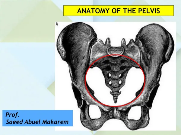
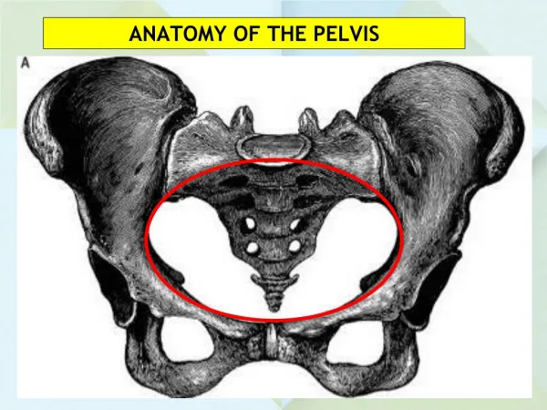
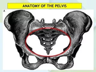
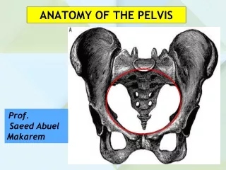
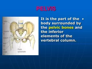
Intro & Definitions • Pelvic Brim (green line) • Imagine a line drawn between promontory of the sacrum, arcuate line of the ilium, pectineal line (pectin of pubis) and pubic crest. • Greater (False) pelvis • All of the bony pelvis ABOVE pelvic brim • Lesser (True) pelvis • All of pelvis BELOW pelvic brim.
Bones et al Bones • Ilium (one on each side): crest, anterior superior and inferior iliac spines and greater sciatic notch. • Pubic bone (one on each side): lesser sciatic notch, tubercle and symphysis • Ischium (one on each side): lesser sciatic notch, spine and tuberosity • Sacrum: foramina for spinal nerves • Coccyx
More bones • Vertebral column: 5 fused sacral and 3-5 fused coccygeal vertebrae • Ilium, pubic bone and ischium meet to form acetabulum for hip joint • Obturator foramen made by articulation of ischium with pubic bone Ilium Pubic bone Ischium
Ligaments • Anterior longitudinal: runs down entire vertebral column. Prevents hyperflexion • Inguinal ligament: arched fibres of external oblique • Pubic Symphysis: secondary cartilaginous joint • Sacroiliac joints anteriorly • Posterior: sacrotuberous, posterior sacrospinous and sacrospinous • Ligaments provide strengthand stability of hip
Male Vs Female Bones: pelvis taller, narrower and more compact. Evolutionary optimised for bipedal locomotion. Acute angle between pubic rami (70 degrees). Contents: rectum, bladder, prostate, anus and male reproductive organs Bones: wider and broader with larger inlet. Optimised for childbirth without compromising bipedal locomotion. Wide angle between pubic rami (100 degrees). Wider acetabulum. Contents: rectum, bladder, anus and female reproductive apparatus
Muscles pubococcygeus • Greater Pelvis: • QuadratusLumborum: from iliac crest to insert into 12th rib and L1-4. Lateral flexor • Psoas Major: from lumbar veterbrae to lesser trochanter of femur. Hip flexor. • Iliacus: from internal iliac fossa to lesser trochanter. Joins with Psoas major = ILIOPSOAS (hip flexor and trunk flexor) • Piriformis : from greater sciatic notch and anterior sacrum to greater trochanter. Lateral rotator • Lesser Pelvis: • MUSCLES CONTROL SPHINCTERS • Diaphragm: pubococcygeus, coccygeus, puborectalis, (pubovaginalis) and illiococcygeus • Levtorani = a sling made by puborectalis, pubo- and ilio-coccygeus. Prevents incontinence. • Sphincter urethrae • (Sphincter prostatae) • External anal sphincter • pubovaginalis elevates vagina iliococcygeus coccygeus Psoas major piriformis iliacus
Vasculature: Arteries • Gonadal artery (branch of abdo aorta, origin L2) • Internal Iliac (anterior + posterior divisions): • Superior vesical • Inferior vesical (vaginal artery in female) • Middle and inferior rectal (superior rectal from inferior mesenteric) • Inferior and superior gluteal • Uterine (uterus, vagina, ureter) • Internal Pudendal (perineum, penis and urethra)
veins • Veins from pelvis follow arteries • Drain to IVC (common iliac joins at L5) • Left testicular drains to left renal not directly into IVC
lymphatics • Lateral pelvic drain everything EXCEPT: • Para aortic drain: gonad + fallopian tube + uterus + ureter • Inferior mesenteric drain: upper rectum • All ultimately drain into lymphatic duct and cisterna chyli
Nerves • Dermatomes: T12 (suprapubic), L1 (groin), L2 (upper thigh), S1, 2, 3, 4, 5 (buttocks, perineal and perianal). S1, 2 (genitals). • Sympathetic: from lumbo-sacral trunk (L1-S5). • Parasympathetic: S2-4 • Lumbar plexus: L1-5 roots lie on Psoas M. Branches: • 3 lateral to Psoas (lateral cutaneous nerve, iliohypogastric, ilioinguinal and • 1 anterior to Psoas: genitofemoral • 2 medial to psoas: femoral, obturator • Sacral Plexus: S1-4 • Pudendal: S2-4. mixed sensory/autonomic • Coccygeal
Autonomic Sympathetic Parasympathetic Pudendal nerve: mixed autonomic and sensory. S2-4 Pelvic splanchnic nerves: preganglionic fibres from S2-4 travel to hypogastric plexus ad from there nerves travel to and synapse at viscera. Cause erection and sphincter relaxation for micturition/defaecation • Hypogastric nerves: preganglionic fibres travel to hypogastric plexus and synapse there then travel to viscera as hypogastric nerves. • Sacral splanchnic nerves: fibres synapse at sympathetic chain and postganglionic fibres travel to hypogastric plexus as a splanchnic nerve.
Divisions of Lumbar plexus lateral cutaneous nerve: sensory to lateral thigh Iliohypogastric: motor to transversus and internal oblique, sensory to mon pubis Ilioinguinal: motor to internal oblique, transversus and conjoint tendon. Sensory to upper medial thigh, labia majora, scrotum and root of penis Genitofemoral: motor to cremaster. Sensory to scrotum, anterior thigh, spermatic fascia and tunica vaginalis. Femoral (L2,3,4): motor to iliacus, pectineus and quadriceps femoris. Sensory to anterior thigh. Obturator :
Sacral Plexus • Formed by L4, 5, S1-5 • Lies on piriformis • Branches: • 6 nerves from sacral roots • Nerve to piriformis • Posterior femoral • Perforating cutaneous • Perineal branch to levatorani • Pelvic splanchnic • Pudendal • Anterior division: • Nerve to Quadratusfemoris • Nerve to Obturatorinternus • Tibial branch of sciatic nerve • Posterior division: • Superior gluteal • Inferior gluteal • Common peroneal branch of sciatic nerve
Pudendal Nerve • Somatic and autonomic • Origins S2-4 • Exits through greater sciatic foramen and re-enters pelvis via lesser sciatic foramen • Travels with pudendal vessels along ischiorectalfossa in Alcock’s canal • Supplies sphincters and genitalia via perineal, dorsal root of penis/clitoris and inferior anal nerves • Promotes ejaculation, sexual arousal, anal and bladder sphincter control.
Coccygeal Nerve • 31st spinal nerve • Forms coccygeal plexus with S5 • Coccygeal plexus gives rise to annococcygeal nerve which supplies sacroccygeal joint and skin over coccyx.
Organs • RENAL TRACT: • Ureters: • Originate at renal hilum at L2 • Path initially medial to vertebrae and at pelvic brim take infero-posterior path • Oblique entry into bladder avoids urinary reflux • Crossed by gonadal artery in pelvis • Posterior to it are psoas and genitofemoral nerve • Under it are uterine artery and vas deferens • Arterial supply via gonadal, renal, vesical, vaginal and aortic branches • Autonomic innervation • Bladder: • Trigonal structure. • Wall has 3 layers of smooth muscles: inner circular and middle/outer longitudinal layers • Arterial supply from superior and inferior vesicalnerves: sympathetic closes bladder neck whilst parasympathetic relaxes detrusor muscle to allow for miturition
Rectum and anus • Rectum • Columnar epithelium • Superior 1/3 covered by peritoneum anteriorly and laterally, middle 1/3 anterior peritoneum only and inferior 1/3 bare • Arteries: superior rectal from inferior mesenteric and middle rectal from internal iliac +inferior rectal from pudendal artery • Veinous drainage from internal venous plexus which drains to: • superior rectal which then drains to inferior mesenteric vein, middle rectal which drains to internal iliac vein and inferior rectal vein which drains into pudendal vein • Anus: • Starts at anorectal junction aka dentate line • Squamous epithelium continuous with skin gradually transforming to columnar as rectum approached • External anal sphincter is skeletal muscle with somatic innervation thus voluntary • Internal anal sphincter is smooth muscle and under autonomic control
Female pelvic viscera • Uterus: • Held at lateral walls by double fold of peritoneum aka broad ligament • Uterine artery • Sympathetic and parasympathetic innervation from pelvic plexus • Venous plexus drain to rectal and vesical veins • Ovaries: • Attached to posterior aspect of broad ligament • Ovarian artery • Right ovarian vein drains to IVC whilst left to left renal vein • Sympathetics from aortic plexus and parasympathetics from pelvic plexus • Fallopian tubes: • Run in free edge of broad ligament • Ovarian and uterine arteries • Vagina: • Opens into vaginal vestibule • Vaginal artery • Sympathetic supply from pelvic plexus and somatic sensory innervation from ilioinguinal and pudendal nerves • Venous drainage from pelvic floor plexus to internal iliac • Clitoris: • Female equivalent of penis • Nerve supply via pudendal
Male pelvic viscera • Scrotum: • layers are skin, dartos muscle, external spermatic fascia, cremaster muscle, internal spermatic fascia, tunica vaginalis and tunica albuginea • Testis: • Testicular (gonadal artery) • pampiniform plexus drain to testicular veins • Testicular vein drains to IVC on right and left renal artery on left • Prostate: • Multi-lobar (5) with posterior groove. Apex at the bottom and base at top • Smooth muscle • Entered by the vasa deferens and seminal vesicals • Contains prostatic urethra • Arterial supply from inferior vesical, middle rectal and occasionally pudendal arteries • Drains to venous plexus and then to internal iliac vein • Sympathetic nerves promote ejaculation and smooth muscle contraction whilst parasympathetics promote erection • Penis and Urethra: • Pre-prostatic, prostatic, membranous and penile urethra • Receives ejaculatory ducts, bulbourethral and urethral glands • Arterial supply from urethral artery, deep artery to penis and dorsal artery of penis • Drainage via superficial and deep dorsal veins of penis • Nerves are sympathetic and parasympathetics for ejaculation and erection. Sensory supply to skin and glans of penis from pudendal nerve
Special places • Inguinal canal: • 4 cm long running from Anterior superior iliac spine and pubic tubercle. • Contains spermatic cord (or round ligament) and ilioinguinal nerve. • Spermatic cord contains: 3 structures (vas deferens, cremaster muscle and pampiniform plexus), 3 arteries (artery to vas, artery to cramster and testicular artery) and 3 nerves (sympathetic, parasympathetic and genitofemoral) • Floor: fibres of external oblique = inguinal ligament • Roof: transversusabdominis and internal oblique • Anterior: external oblique and internal oblique • Posterior: transversalis fascia and conjoint tendon • Alcock’s canal: • Where pudendal nerve, vein and inetrnalpudendal artery run. • Formed by obturatorinternus fascia • Runs on the lateral wall of ischiorectalfossa • Femoral canal: • Contains lymphatic vessels and cloquet’s lymph node • Anterior border is inguinal ligament • Posterior border is pectineal ligament • Medial border is lacunar ligament • Lateral border is femoral vein • Site of bowel herniation • Pubic tubercle: • Herniae above ad medial are inguinal and those below and lateral are femoral
Quiz Q1) On the bony pelvis: • A) true pelvis lies between iliac crests T/F • B) the acetabulum formed by contributions from all parts of hip bone T/F • C) male pelvic inlet more oval than female in shape T/F • D) angle between pubic rami wider in male T/F • E) pelvic out let is between symphysis pubis and sacral tuberosity T/F
Quiz Q1) On the bony pelvis: • A) true pelvis lies between iliac crests T/F • B) the acetabulum formed by contributions from all parts of hip bone T/F • C) male pelvic inlet more oval than female in shape T/F • D) angle between pubic rami wider in male T/F • E) pelvic out let is between symphysis pubis and sacral tuberosity T/F
Q2) Levator Ani • A) has fibres which assist continence by pulling rectum backwards T/F • B) lies inferior to ischiorectal fossa T/F • C) is supplied by anterior rami of S1-2 T/F • D) Contracts during defaecation T/F
Q2) Levator Ani • A) has fibres which assist continence by pulling rectum backwards T/F • B) lies inferior to ischiorectal fossa T/F • C) is supplied by anterior rami of S1-2 T/F • D) Contracts during defaecation T/F
Q3) On sphincters of the anus • A) the anus contains longitudinal and circular muscle T/F • B) External sphincter composed of involuntary muscle T/F • C) external sphincter continuous with muscle of rectum T/F
Q3) On sphincters of the anus • A) the anus contains longitudinal and circular muscle T/F • B) External sphincter composed of involuntary muscle T/F • C) external sphincter continuous with muscle of rectum T/F
Q4) on the inguinal canal: • A) contains spermatic cord and splanchnic nerve T/F • B) posterior border is transversus abdominis and internal oblique T/F • C) floor is inguinal ligament T/F • D) carries round ligament in females T/F
Q4) on the inguinal canal: • A) contains spermatic cord and splanchnic nerve T/F • B) posterior border is transversus abdominis and internal oblique T/F • C) floor is inguinal ligament T/F • D) carries round ligament in females T/F
Q5) on origins of nerves • A) lumbar plexus from L1-5 T/F • B) pudendal arises from S2-4 T/F • C) parasympathetic plexus arises from S2-4 T/F • D) hypogastric nerves carry postganglionic fibres T/F
Q5) on origins of nerves • A) lumbar plexus from L1-5 T/F • B) pudendal arises from S2-4 T/F • C) parasympathetic plexus arises from S2-4 T/F • D) hypogastric nerves carry postganglionic fibres T/F
Fin • Useful books: • Instant anatomy: good for surface anatomy, blood vessels and nerves • Netter’s atlas • (Anatomy recall) • Websites: • www.iwanttobeasurgeon.com (down for construction at present) • www.iwanttobeasurgeon.blogspot.com • www.instantanatomy.net • Apps: • Gray’s anatomy (2 quid!) • Netter’s flash cards (twenty pounds but useful for revision on tube) • DVD: • Acland’s (AMAZING and free from Warwick University website or youtube. £130 for DVD set) • Exam Material: • Pastest has over 800 anatomy questions and even more useful when you sit finals Have to pay though :( • http://ect.downstate.edu/courseware/haonline/quiz/practice/u7/quiztop7.htm (excellent for uestions on cadaveric dissections)