The Pelvis
The Pelvis. ANHB 2212 – 2008 Avinash Bharadwaj. Bony Pelvis. Hip bones Sacrum Joints and ligaments. Hip Bone. Ligaments. Sacrospinous. Sacrotuberous. Notches Foramina. Other ligaments. The walls. Pelvis as a container Muscles Piriformis Obturator muscles (Internus/externus)

The Pelvis
E N D
Presentation Transcript
The Pelvis ANHB 2212 – 2008 Avinash Bharadwaj
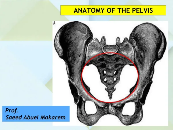
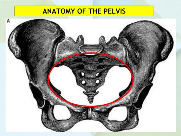
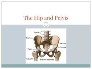
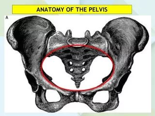
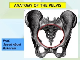
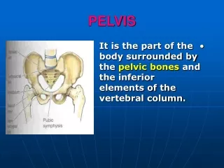
Bony Pelvis • Hip bones • Sacrum • Joints and ligaments
Ligaments • Sacrospinous • Sacrotuberous • Notches Foramina • Other ligaments
The walls • Pelvis as a container • Muscles • Piriformis • Obturator muscles (Internus/externus) • Membrane • Vessels and nerves
The Floor • Pelvic diaphragm • Pubis sacrum (coccyx) • Obturator fascia
The Floor • Pelvic diaphragm • Levator ani • Puborectalis • Pubococcygeus • Iliococcygeus • Coccygeus (ischio-) • Perineum • Urogenital region • …diaphragm • Anal region
Urogenital Region • Male urethra • Penis • Corpus spongiosum and corpora cavernosa • Muscles • Comparable female structures
The Pelvis – “True” and “False” • Iliac fossae • The pelvic brim • Pelvic cavity
Contents • Urinary system • Bladder and urethra • Repoductive system • Male / female • Digestive system • Rectum – anal canal • Peritoneum…
Urinary Bladder • Muscular wall • Reservoir • Sphincters • Internal – smooth • External – urogenital diaphragm - skeletal • Variation in size, shape and location… • Empty – Tetrahedral, Pelvis • Full – ovoid, partly abdominal
Urogenital Organs - Male • Prostate • Seminal vesicles • Ductus deferens
Female Reproductive Organs • Uterus • Fundus, body, cervix • Vagina • Angles • Fornix • Ovary and the tube
Female Reproductive Organs • Uterus • Fundus, body, cervix • Non-pregnant • Pelvic • Pregnant • Pelvic 12 weeks • Abdominal • 20 weeks – umbilicus • 36 weeks - xiphisternum
Female Reproductive Organs • Ovaries • Descent • Stops in the pelvis • Ligaments • Round ligament • Ovarian artery • From aorta
Female Reproductive Organs • Uterine tubes • Parts • Also note • Ovarian artery • Uterine artery • Ligaments
Rectum and Anal Canal • The junction • Developmental correlation • Venous anastomoses • Nerves and lymphatics • Mucosa and sphincters Internal sphincter – smooth muscle External sphincter - skeletal
Rectum and Anal Canal • Lower part • Ectodermal • Systemic veins • “Modified skin”
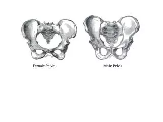
The Pelvis – Birth Canal • Is it adequate? • How do we know? • Sexual dimorphism • Variations and types
Pelvic Dimensions • Inlet • Midcavity • Outlet • A-P • Transverse • Oblique
Types of Female Pelvis • Gynaecoid • Android • Antrhopoid • Platypelloid • Abnormalities