Radiology Case Studies: Pelvic Imaging Analysis with Clinical Correlations
220 likes | 249 Vues
Explore detailed radiology cases illustrating pelvic imaging findings in female patients with pelvic pain. Identify structures, imaging sequences, and make differential diagnoses.

Radiology Case Studies: Pelvic Imaging Analysis with Clinical Correlations
E N D
Presentation Transcript
Case 1 36 year old woman with pelvic pain
What type of images are these (CT, MRI or plain films)? • What plane are they in (axial, sagittal or coronal)? • What type of imaging sequence is this, T1 or T2? • Hint, look at the fluid in the bladder is it bright or dark. Remember that fluid is bright on T2 weighted sequences
On the this image, there are 3 soft tissue structures in the pelvis, two you should be able to identify and one you shouldn’t. What are the 2 normal structures you see in this female patient? Uterus Bladder ???????
Axial This is a T1 weighted sequence • What imaging plane is this? • What imaging sequence is this, T1 or T2? Hint, look at the subcutaneous fat, is it bright or dark (fluid is also dark, not shown)? What other things are bright on T1 weighted images. Fat, blood and MRI contrast are bright on T1 weighted sequences
Now for the diagnosis in this case. Do you see another structure that is high signal on these T1 weighted images? Look in the left side of the pelvis. (click for arrow ) We then used an MRI trick to help us figure out what was causing this structure to be bright on T1 weighted images.
We use a fat suppression technique, all of the fat is dark, even the fat in the bone marrow. This helps us determine the cause of the high signal structure in the left ovary. So what do you think is causing this lesion to stay bright on this fat suppressed T1 weighted sequence???? • What is the difference between these 2 images? Hint, look at the fat.
Since this high signal did not get dark with fat-suppression then it must be getting its high signal from blood products as we didn’t give contrast. This is an endometrioma, an area of endometrial tissue, similar to the lining of the uterus but not in the uterus (ectopic in location). This can be a cause of pelvic pain in female patients.
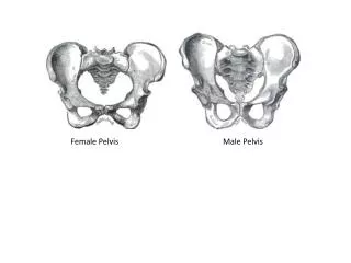
What type of image is this? • Can you point to the different bones and joints that make up the bony pelvis (ilium, pubic rami and sacrum, SI joints and pubic symphysis).
Is there anything that looks “funny” about any of these joints? • Hint look at the pubic symphysis and the SI joints
What do you think could have caused this (trauma, congenital defect or infectious disorder)? Click for answer
For extra credit: do you see any fractures of the bones of the pelvis? If you see this you, have a bright career in radiology (click for answer).
CT Soft tissue window • What time of images are these? (Plain Films, CT or MRI). • What type of window is this set for (soft tissue or bone)?
This is a bone window, note how you can see the bone detail between the cortex and marrow space.
Is this a male or a female patient? Look at the structures behind the bladder. Does this look like a uterus or could these be seminal vesicles? These are seminal vesicles
What is the structure just posterior to the seminal vesicles? Hint, it has air in it. Rectum
What are the small round structures just anterior to the iliopsoas muscles? Hint, they are tubular, bilateral, symmetric and on every image. External iliac artery and vein
Take a look at the bladder, what is the round low attenuation area (dark area) in it? Foley catheter
You see contrast in 2 places in these images, in the bladder and anterior to the bladder. What is the name anatomic space anterior to the bladder? Prevesicle space
Hint, is the prevesicle space intra or extraperitoneal? • This is a bladder rupture in a trauma patient. There are 2 types of bladder ruptures, intraperitoneal and extraperitoneal. For extra credit, what type is this? This is an extraperitoneal bladder rupture. For an intraperitoneal bladder rupture you would see contrast between the bowel loops. There is a big difference in treatment between the two types of rupture. Intraperitoneal ruptures are treated surgically where extraperitoneal ruptures are treated conservatively.