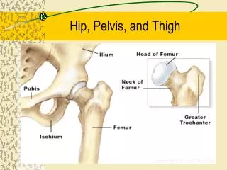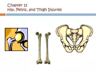
Hip and Pelvis
This chapter focuses on critical radiographic techniques for imaging the hip and pelvis. Key aspects include proper facility identification, correct marker placement, avoidance of artifacts, and optimal film size (10x12). Important parameters such as density and contrast are highlighted, including a recommended kVp range of 75-85. The text emphasizes the significance of visualizing bony structures and soft tissues in an AP projection and includes techniques for detecting rotation and ensuring complete anatomical representation. Specific imaging methods like Hip AP, Frog-leg lateral, and Axiolateral views are discussed in detail.

Hip and Pelvis
E N D
Presentation Transcript
Hip and Pelvis Chapter 6
Hip AP • Facility Identification • Correct Marker Placement • No Preventable Artifacts • Correct Film Size (10 x 12 lw unless indicated)
Hip AP • Density • Controlled by mAs • Overall the density is not too dark or too light • Contrast • Optimal kVp 75-85 • Bony trabecular patters and cortical outlines visualized as well as soft tissues (fat pads) of the hip are demonstrated
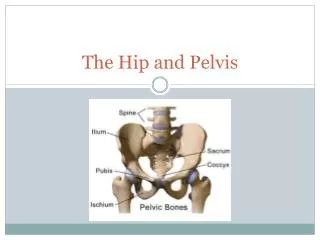
Hip AP • Pelvis is in true AP projection • The ischial spine is aligned with the pelvic brim • The sacrum and coccyx are aligned with the symphysis pubis • The obturator foramen is open
Hip AP • Detecting rotation toward the affected hip • If the ischial spine is demonstrated without pelvic brim superimposition • The sacrum and coccyx are rotated away from the affected hip • The obturator foramen is narrowed
Hip AP • Detecting rotation away from the affected hip • The sacrum and coccyx are rotated toward the affected hip • The ischial spine is closer to the acetabulum • The obturator foramen is widened
Hip AP • The femoral neck is demonstrated without foreshortening • The greater trochanter is demonstrated in profile laterally • The lesser trochanter is superimposed by the femoral neck
Hip AP • The femoral head and acetabulum are in the center of the collimated field • Any orthopedic appliance are located at the hip are included in their entirety.
Hip AP • Gonadal shielding should be used on all males and females if it may be placed so that anatomy is not obstructed
Hip Frog leg Lateral • The pelvis is in a true AP position • The ischial spine is aligned with the pelvic brim • The sacrum and coccyx are aligned with the symphysis • The obturator foramen is open
Hip Frog leg Lateral • The lesser trochanter is demonstrated in profile medially • The femoral neck superimposes the greater trochanter • The neck if foreshortened and is ½ way between the femoral head and the lesser torchanter
Hip AxiolateralDanielus Miller • Placing lead flat contact shields over the unused portion of the cassette will reduce scatter and help improve contrast
Hip AxiolateralDanielus Miller • The femoral neck is demonstrated without foreshortening • The greater and lesser trochanters are about the same transverse level
Hip AxiolateralDanulus Miller • If the angle formed between the femur and the central ray is too large, the trochanter is demonstrated proximal to the lesser trochanter and is superimposed by a portion of the femoral neck • If the angle is too small, the greater trochanter is demonstrated distal to the lesser trochanter. (this seldom occurs due to table top)
Pelvis AP • Pelvis is demonstrated without rotation • The ischial spines are aligned with the pelvic brim • The sacrum and coccyx are aligned with the symphysis • The ilia and the obturator foramina are uniform in size and shape
Pelvis AP • Male –vs- Female Pelvis • Male pelvis is more heart shaped • The obturator foramina and acetabula are larger and more bulky • Female pelvis is more oval
Pelvis AP • The femoral neck is demonstrated without foreshortening • The greater trochanter is demonstrated in profile laterally • The lesser trochanter is superimposed by the femoral neck
Pelvis AP • Detecting rotation toward the affected hip • If the ischial spine is demonstrated without pelvic brim superimposition • The sacrum and coccyx are rotated away from the affected hip • The obturator foramen is narrowed
Pelvis AP • Detecting rotation away from the affected hip • The sacrum and coccyx are rotated toward the affected hip • The ischial spine is closer to the acetabulum • The obturator foramen is widened
Pelvis AP • All anatomy is included on the film • Inferior sacrum is in the center of the film • The ilia, symphysis ischia acetabulum, femoral necks and heads, greater and lesser trochanter are included on film