Download
1 / 19
190 likes | 365 Vues
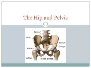
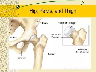
Hip, Pelvis and Distal Femur. Things to know for Pelvis. Cassette Size 14 x 17 crosswise One view AP 80 @ 12 or 90 @ 8 No shielding No collimation Marker. Positioning for AP Pelvis. Patient is supine on table, Arms at side Align the midsagittal plane to the center of table

E N D
Things to know for Pelvis • Cassette Size 14 x 17 crosswise • One view AP • 80 @ 12 or 90 @ 8 • No shielding • No collimation • Marker
Positioning for AP Pelvis • Patient is supine on table, Arms at side • Align the midsagittal plane to the center of table • Make sure pelvis is not rotated • Separate feet slightly and internally rotate feet 15 to 20 degrees.
Central Rayis midway between pubis level of the ASIS and the Symphysis SID 40
Radiographic critique • Pelvic girdle, L5, sacrum and coccyx • Femoral heads, neck and greater trochanters • Pelvic Wings symmetrical
Hip • 2 Views. AP and Lateral • 10 x 12 cassette • Measures 17 • 80 @ 12 • Shield • Mark
Positioning for AP Hip • 10 x 12 lengthwise • Patient supine on table • Mid-line of femoral neck to mid-line of table • Rotate leg 15-20 degrees internally • make sure no rotation
Central ray- 3-4 inches below ASIS and 1-2 inches medially • SID 40
Radiographic critique • 1/3 of femur • Acetabulum and adjacent pubis, ischium, and ilium • Greater trochanter
Lateral Hip • 10 x 12 crosswise • Patient supine • Flex knee of injured leg with the sole of foot against the knee of opposite leg • Abduct femur 45 degrees • Center mid-femoral neck to IR
Central ray mid-femoral neck SID 40
Critique • A lateral view of acetabulum, femoral neck • Lesser trachanter
Distal Femur • 2 views AP and Lateral • 14 x 17 cassette lengthwise • measures 13 • 75 @ 12 • Shield • Mark
Positioning for AP • Patient supine • femur to mid-line of table • Rotate leg about 5 degrees internally to ensure true AP • Include knee joint
Critique • Distal 2/3 of femur • Knee joint • Condyle should appear symmetrical
Lateral Distal Femur • Roll patient upon affected side so injury will be closer to IR • Flex knee 45 degrees • Move uninjured leg out of the way • Align femur with mid-line of table • Include knee joint
Critique • Distal 2/3 of femur • Included knee joint • Condyles should be superimposed.