Download
1 / 51
1.08k likes | 3.09k Vues
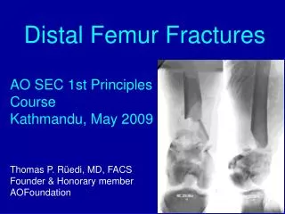
Distal Femur Fractures. AO SEC 1st Principles Course Kathmandu, May 2009. Thomas P. Rüedi, MD, FACS Founder & Honorary member AOFoundation. Albin Lambotte 1886- 1955 Pioneer and genius of modern operative fracture treatment. ....already 100 years ago recognized that articular

E N D
Distal Femur Fractures AO SEC 1st Principles Course Kathmandu, May 2009 Thomas P. Rüedi, MD, FACS Founder & Honorary member AOFoundation
Albin Lambotte 1886- 1955 Pioneer and genius of modern operative fracture treatment..... ....already 100 years ago recognized that articular fractures must be fixed rigidly with plates and screws in order to allow for early, pain free motion
Unfortunatley, Lambottes’ recommendations were forgotten or his superb operative technique and soft tissue care could not be reproduced, so that X-rays like these can be seen until today: Such attempts at surgery are totally inadequate !!!!
1965 Allgöwer fixed this IIIB open, 33 C1 fx from hunting incident as an emergency with 95° blade plate and secondary bonegraft. • at 6 mo bony union, limited flexion (recurvatum), no pain • 25 years later: acceptable function, no signs of osteoarthritis
requires careful planning What are the challenges ? • complex anatomy of knee / ligaments • high energy / polytrauma • short distal segment • & long leever arm • positioning, approach • soft tissue cover • choice and purchase • of implant in bone • functional after care
Planning of surgery • correct diagnosis for classification • condition of patient as a whole • soft tissue conditions of injured area • timing and sequence of surgery • step-by-step: positioning, approach, reduction, • preliminary fixation, choice of implant • any minimally invasive technique must be • decided beforehand and carefully performed
A extraarticular / supracondylar B intra-articular unicondylar ( incl.Hoffa) A B C C intra-articular bicondylar Classification (Müller AO) 33 - • Often high energy, • open fractures, • neuro-vascular • injuries in 3-4%
W.G.m, 19y: collision with ratrac while skiing : • Cranio-facial fractures, GCS 9 ISS 38 • Hemo-pneumothorax, rib fractures • 23- C2 fracture left distal humerus, bilateral distal radius fx. • Floating knee with II° open distal femur and tibia fracture, neuro- • vascular intact
W.G.m, 19y: collision with ratrac while skiing : ISS 38 • Emergency fixation:- DCS for distal femur • lag screw for tibia plateau and joint bridging ext. fix. • ORIF dist. humerus • After 10 days: • lateral bridge plate for tibia, • ex-fix as reduction aid • - Fixation of both radius • fractures - physiotherapy
Due to slow healing of tibia: Cancellous autograft after 5 months • Good functional result after one year, slight varus back to work and sports 12 mo 5 mo W.G.m, 19y: collision with ratrac while skiing : ISS 38
Distal femur fracture: choice of implants • Classical: • 95° angle blade plate • DCS: dynamic condylar screw • condylar buttress plate • New: • LISS: less invasive stabilisation system • locking condylar buttress plate • LCP: locking compression plate • retrograde im-nail
Sch.W. 61y,m MVA: distal Femur fracture 33- C, circumferential degloving of whole leg case of P.Tondelli Emergency ORIF with 95° angled blade plate, Harvesting of defattened, degloved skin for later use
Sch.W. 61y,m MVA: distal Femur fracture 33- C, circumferential degloving of whole leg case of P.Tondelli Secondary split skin graft with defattened skin Satisfactory functional result
Patient positioning: • knee flexed 30- 45° to reduce • pull of gastrocnemius muscle • radiolucent table Approaches: Para-patellar lateral
Hunting accident:III-C open (artery,vein + nerve), 33-C3 fract, Control angiogram 1) Preliminary fixation with DSC and 3cm shortening 2) Repair of politeal artery & vein with venous grafts, nerve bruised, but intact 3) Completion of ORIF and compartment release
Hunting accident:III-C open(artery, vein+ nerve), 33-C3 fract. at 3 months: good function, uneventful healing, weightbearing w. 40 kg to correct 3cm shortening: proximal one step lengthening, good one year result
6 weeks after injury postop R.O.m, 44y: polytrauma w. 33-C3 open fx After 6 we in traction >> 4cm shortening, mal alignment, stiff joints Plan: indirect reduction w distractor, minimal exposure, DCS
27 weeks 4 years R.O.m, 44y: polytrauma w. 33-C3 open fx Intensive postop. Physiotherapy > return of function. Good healing Removal of sequestrum, playing tennis after 2y, follow-up at 4y
A.B.m 26y, motorbike injury, III B open, 33- C3 fracture neuro-vascular intact Emergency ORIF, attempt at anatomical reconstruction of condyles, fixation w. condylar buttress plate, all stripping of the bone is traumatic such surgery can hardly be done through a keyhole incision
A.B.m 26y, motorbike injury, III B open, 33- C3 fracture In spite of considerable exposure, bone graft & cerclage wire, unproblematic healing, return of satisfactory function, here at one year follow-up
Similar 33- C3 fracture in 70 y old man, Initial ORIF w condylar buttress plate, osteoporosis, poor purchase, no support Collapse of fixation after 5 we redo w 95° angle blade plate: redo w 95° angle blade plate, bony union after 1 y, satisfactory functional result 5 weeks 1 year
With the new concept of the,internal fixator based onangular stability of the screws, such screw loosening & collapse will not occur anymore! LISSl Less invasive stabilisation system
LISS(less invasive stabilisation system) • reduction: - direct (vision) of articular components • - indirect of meta- / diaphysis • minimally invasive, submuscular insertion of • long bridging plates • locking head screws provide angular stability • - uni-cortical or bi-cortical • - plate not pressed against bone • excellent purchase also in osteopenic bone, • - eg. periprosthetic fractures • no bone graft required
Distal femur fracture 33- C2 (metaphyseal comminution) • secondary ORIF with LISS • reconstruction and alignment of articular surface/ block > plate insertion postop 2 mo • initial, temporary joint bridging external fixator,
Step-by-step procedure for the LISS • Planning position / length of LISS • Submuscular insertion of plate sliding along femur • Preliminary distal fixation for indirect reduction with distractor • Lateral view and ap after reduction
LISS or LCP Internal fixator principle DCU LISS Peri-prosthetic fractures or in osteopenic bone: • poor purchase of standard • screws / implants • locking head screws provide • - angular stability • - less risk for pull- out Combi-hole= LCP
Advantages of LISS in periprosthetic fractures: • Locking head screws - providing firm purchase in osteoporotic bone • - unicortical application (around stem of prosthesis) • no cement required
LCP: clinical handling tests (2000) Dr.Ch.Sommer, Chur I.K., 40y, distal femur C2-type, 2°open
LCP: clinical handling tests (2000) Dr.Ch.Sommer, Chur I.K., 40y, distal femur C2-type, 2° open
3months LCP: clinical handling tests (2000) Dr.Ch.Sommer, Chur I.K., 40y, distal femur C2-type, 2° open
6 months LCP: clinical handling tests (2000) Dr.Ch.Sommer, Chur I.K., 40y, distal femur C2-type, 2° open
left right M.36 y. Motorbike accident, polytrauma(case of Dr.Turchetto) - abdominal injuries (spleen & ileum) - bilateral identical, segmental distal 3rd femur fractures • preliminary external fixation until recovery >> bilat. retrograde im-nail
rigth M.36 y. Motorbike accident, polytrauma - bilateral identical segmental femur fract. • secondary bilateral retrograde nailing : • Right side: • - ORIF intraarticular fx w cancellous screw • - retrograde nail insertion
left M.36 y. Motorbike accident, polytrauma - bilateral identical segmental femur fract. • Left side: • percutaneous reduction / cannulated screw • minimal approach for retrograde nail
M.36 y. Motorbike accident, polytrauma • bilateral identical segmental femur fract. • 7 days postoperatively (case of Dr.Turchetto)
M.36 y. Motorbike accident, polytrauma bilateral identical segmental femur fract. • 40 days follow up >> callus formation right left (case of Dr.Turchetto)
Conclusions: • distal femur fractures 33- A- C are: • - absolute indications for ORIF • - often high energy injuries, open, & polytrauma • require careful planning as to: • - timing, positioning, approaches (soft tissues !) • - reduction techniques • - choice of implant • variety of implants today available: • - 95°angle blade pl. / DCS condylar buttress pl. • - LISS / LCP (locking head screws > angular stability) • - retrograde intra-medullary nails • minimally invasive techniques w indirect reduction and • bridging implants to be preferred
Motorcycle injury: 33- C3 (not suited for blade plate or DCS) ORIF w. condylar buttress plate, uneventful healing and functional recovery