Download
1 / 78
1.16k likes | 2.24k Vues
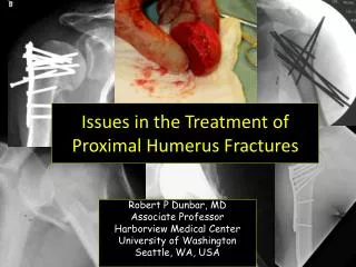
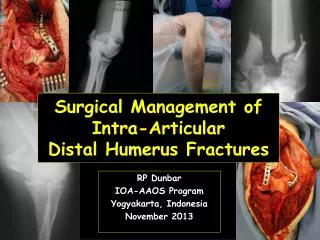
Fractures of the Distal Humerus. Laura S. Phieffer, MD Original Authors: Jeffrey J. Stephany, MD and Gregory J. Schmeling, MD; March 2004 New Author: Laura S. Phieffer, MD; Revised January 2006. distal humeral triangle. Functional Anatomy.

E N D
Fractures of the Distal Humerus Laura S. Phieffer, MD Original Authors: Jeffrey J. Stephany, MD and Gregory J. Schmeling, MD; March 2004 New Author: Laura S. Phieffer, MD; Revised January 2006
distal humeral triangle Functional Anatomy • Hinged joint with single axis of rotation (trochlear axis) • Trochlea is center point with a lateral and medial column
Functional Anatomy • The distal humerus angles forward • Lateral positioning during ORIF facilitates reconstruction of this angle
Surgical Anatomy • The trochlear axis compared to longitudinal axis is 94-98 degrees in valgus • The trochlear axis is 3-8 degrees externally rotated • The intramedullary canal ends 2-3 cm above the olecranon fossa
Surgical Anatomy • Medial and lateral columns diverge from humeral shaft at 45 degree angle • The columns are the important structures for support of the “distal humeral triangle”
Mechanism of Injury • The fracture is related to the position of elbow flexion when the load is applied
Evaluation • Physical exam • Soft tissue envelope • Vascular status • Radial and ulnar pulses • Neurologic status • Radial nerve - most commonly injured • 14 cm proximal to the lateral epicondyle • 20 cm proximal to the medial epicondyle • Median nerve - rarely injured • Ulnar nerve
Evaluation • Radiographic exam • Anterior-posterior and lateral radiographs • Traction views are necessary to evaluate intra-articular extension and for pre-operative planning (“ligamentotaxis”) • Traction removes overlap • CT scan helpful in selected cases • Comminuted capitellum or trochlea
OTA Classification • Follows AO Long Bone System • Humerus, distal segment given # 13 • 3 Main Types • Extra-articular fracture (13-A) • Partial articular fracture (13-B) • Complete articular fracture (13-C) • Each broad category further subdivided into 9 specific fracture types
OTA Classification • Humerus, distal segment (13) • Types • Extra-articular fracture (13-A) • Partial articular fracture (13-B) • Complete articular fracture (13-C)
OTA Classification • Humerus, distal segment (13) • Types • Extra-articular fracture (13-A) • Partial articular fracture (13-B) • Complete articular fracture (13-C)
OTA Classification • Humerus, distal segment (13) • Types • Extra-articular fracture (13-A) • Partial articular fracture (13-B) • Complete articular fracture (13-C)
Summary - Classifications • Classifications are useful for research! • Classification data may not be reproducible between different surgeons! • Classification data may not be reproduced by the same surgeon at different times!
Summary - Classifications • Meaningful patterns • Extra-articular distal humerus • Medial epicondyle • Lateral epicondyle • Distal metaphyseal humerus • Partial articular distal humerus • Capitellum • Trochlea • Complete articular distal humerus
Group 1 Lateral epicondyle Medial epicondyle Capitellum Trochlea Group 2 Distal humerus Extra-articular Intra-articular Simplicity Note: Fixation tactics & implants are based on groups
Treatment Principles • Anatomic articular reduction • Stable internal fixation of the articular surface • Restoration of articular axial alignment • Stable internal fixation of the articular segment to the metaphysis and diaphysis • Early range of motion of the elbow
Treatment: Open Fracture • Emergent I&D • Definitive reduction and internal fixation • Temporary external fixation across elbow if definitive fixation not possible • Definitive fixation at repeat evaluation • Empiric antibiotic therapy • Repeat evaluation in OR until soft tissue closure (2-5 days)
Treatment: Closed Fracture • Definitive reduction and internal fixation • Timing • Within 24 hours or at 5-7 days • The inflammatory response peaks at 3 days post injury. ORIF during that peak may lead to excessive heterotopic ossification • Empiric antibiotic therapy
Fixation Methods: Group 1 • Lag screw fixation • Comminution is supported by small or mini-fragment buttress plate • Bone graft is considered for comminution and required for bone loss
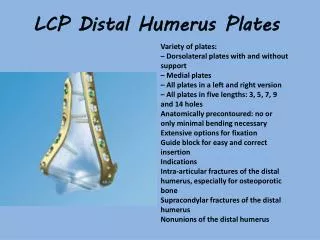
Fixation Methods: Group 2 • Lag screw fixation if possible • Two column plate fixation • Not necessarily at 90 degrees to each other • Bone graft is considered for comminution and required for bone loss • Role of locked plating ?
Literature: Schemitsch, et al, 1994 • Tested 2 different plate designs in 5 different configurations • Distal humeral osteotomy with and without bone contact • Conclusions: • For stable fixation the plates should be placed on the separate columns but not necessary 90 degrees to each other
Literature: Jacobson, et al, 1997 • Tested five constructs • All were stiffer in the coronal plane than compared to the sagittal plane • Strongest construct • medial reconstruction plate with posterolateral dynamic compression plate
Literature: Korner, et al, 2004 • Biomechanically compared double-plate osteosynthesis using conventional reconstruction plates and locking compression plates • Conclusions • Biomechanical behavior depends more on plate configuration than plate type. Advantages of locking plates were only significant if compared with dorsal plate application techniques (not 90/90)
Other Potential Surgical Options • Total elbow arthroplasty • Comminuted intra-articular fracture in the elderly • Promotes immediate ROM • Usually limited by poor remaining bone stock • “Bag of bones” technique • Rarely indicated if at all • Cast or cast / brace • Indicated for completely non-displaced, stable fractures
Literature: John, et al, 1994 • 49 patients (75-90 yrs) • 41/49 Type C • Conclusions • No increase in failure of fixation, nonunion, nor ulnar nerve palsy • Age not a contra-indication for ORIF
Literature: Cobb & Morrey, 1997 • 20 patients • (avg age 72 yrs) • TEA for distal humeral fracture • Conclusion • TEA is viable treatment option in elderly patient with distal humeral Fracture
Literature: Frankle et al, 2003 • Comparision of ORIF vs. TEA for intra-articular distal humerus fxs (type C2 or C3) in women >65yo • Retrospective review of 24 patients • Outcomes • ORIF: 4 excellent, 4 good, 1 fair, 3 poor • TEA: 11 excellent, 1 good • Conclusions: TEA is a viable treatment option for distal intra-articular humerus fxs in women >65yo, particularly true for women with assoc comorbidities such as osteoporosis, RA, and conditions requiring the use of systemic steriods
Surgical Treatment: Group I • Supine with arm on arm board • Sterile tourniquet if possible • Medial or lateral incision • Ulnar nerve transposition considered only if required implants in groove (medial fractures) • As complexity of fracture pattern increases a more extensile exposure should be considered (see Exposures: Group II)
Surgical Treatment: Group I • Fragments are reduced and held with K-wires • Lag screws replace K-wires • Intra-articular screws can be buried in cartilage • Back to front screw direction possible with larger capitellar or trochlear fragments • Small fragment or modular hand plates used to buttress when fracture is comminuted
Surgical Treatment: Group II • Lateral decubitus position • Arm hanging over a post • Sterile tourniquet if possible • Midline posterior incision • Exposure ?
Exposures • Reduction influences outcome in articular fractures • Exposure affects ability to achieve reduction • Exposure influences outcome! • Choose the exposure that fits the fracture pattern
Exposures: Group II • Triceps splitting • Allows exposure of shaft to olecranon fossa • Only indicated for high extra-articular Group II fractures • Extra-articular olecranon osteotomy • Allows adequate exposure of the distal humerus but inadequate exposure of the articular surface • Indicated for extra-articular Group II fractures
Literature: Voor, et al, 1995 • Recommend extra-articular osteotomy over intra-articular osteotomy in all cases • Interesting biomechanical study • Clinical study indicated to justify
Exposures: Group II • Intra-articular olecranon osteotomy • Types • Transverse • Indicated for intra-articular Group II fractures • Technically easier to do • Higher incidence of nonunion (Gainor, et al, 1995) • Olecranon hardware removal in 80% of cases
Exposures: Group II • Intra-articular olecranon osteotomy • Types • Chevron • Indicated for intra-articular Group II fractures • Technically more difficult • More stable • Olecranon hardware removal in 80% of cases
Osteotomy Fixation Options • Tension band technique • Dorsal plating
Osteotomy Fixation • Tension band technique • Anti-shear component • K-wires • Easier to place • Less stable than screw
Osteotomy Fixation • Tension band technique • Anti-shear component • 6.5 mm screw plus washer • Beware of the bow of the proximal ulna, which may cause a medial shift of the tip of the olecranon if a long screw is used. • More stable From Hak and Golladay, JAAOS, 8:266-75, 2000
Osteotomy Fixation • Tension band technique • Tension band • Wire • Dual twist technique • Often palpable necessitating removal • Braided cable • Small crimp less palpable but still can be prominent
Tension band screw Tension band Wire
Osteotomy Fixation • Dorsal plating • Low profile periarticular implants now available allowing antishear screw placement through the plate • No clinical or biomechanical studies yet published using these plates
Chevron Osteotomy • Expose olecranon and mobilize ulnar nerve • If using screw/TBW fixation, pre-drill and tap for 6.5mm screw placement down the ulna canal • Small, thin oscillating saw used to cut 95% of the osteotomy • Osteotome used to crack and complete it
Exposures: Group II • Triceps-sparing postero-medial approach (Byran-Morrey Approach) • Midline incision • Ulnar nerve identified and mobilized • Medial edge of triceps and distal forearm fascia elevated as single unit off olecranon and reflected laterally • Resection of extra-articular tip of olecranon
Exposures: Group II • Triceps-sparing postero-medial approach • An alternative to mobilizing the insertion of the triceps off the olecranon in continuity with the forearm fascia is an extra-articular osteotomy of the olecranon (osteoanconeus flap) • Anterior transposition of ulnar nerve • Triceps re-attached with suture through bone
Surgical Treatment: Group II • Lateral decubitus position • Arm hanging over a post • Sterile tourniquet if possible • Midline posterior incision • Exposure ? • Reduction and K-wire fixation • Lag screws inserted and K-wires removed • Bi-column plating • Reconstruction of triceps insertion per exposure chosen
To transpose or not to transpose? • Identification and mobilization of the ulnar nerve is required for Group II and medial Group I fractures • Ulnar nerve palsy may be related to injury, surgical exposure and mobilization, compression by implant, or scar formation
To transpose or not to transpose? • Wang, et al, in a consecutive series of distal humeral fractures treated with ORIF and anterior ulnar nerve transposition had no post-operative ulnar nerve compression syndrome. Overall results: E/G 75%, F 10%, and P 15%. They conclude that routine anterior transposition indicated.