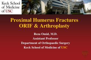
Proximal Humerus
Proximal Humerus. Fractures. Principles of Diagnosis, Decision Making and Treatment. Christopher G. Finkemeier, MD, MBA Revised: May 2011. Acknowledgement: AO faculty lecture archive. Objectives. 1. Learn the principles of diagnosis. 2. Learn the principles of decision making.

Proximal Humerus
E N D
Presentation Transcript
Proximal Humerus Fractures Principles of Diagnosis, Decision Making and Treatment Christopher G. Finkemeier, MD, MBA Revised: May 2011 Acknowledgement: AO faculty lecture archive
Objectives 1. Learn the principles of diagnosis 2. Learn the principles of decision making 3. Learn the various treatment options
Epidemiology All upper extremity fractures 1. forearm fxs 2. proximal humerus fxs All fractures in patients > 65 yrs 1. hip fxs 2. “colles” fxs 3. proximal humerus fxs
HUMERAL HEAD: precarious blood supply AVN GREATER TUBEROSITY: supra/infraspinatus insertion SURGICAL NECK/SHAFT: deltoid/pectoralis major largely dictates fx behavior compression: stable shear: unstable LESSER TUBEROSITY: subscapularis insertion 4 Anatomic Parts Deforming forces determine fx displacement
Vascular Supply • Lateral ascending branch of anterior humeral circumflexartery • Damage may lead to AVN
Gerber et al., JBJS, 1990 Humeral Head Vascularity Non shaded area is supplied by the lateral ascending branch of the anterior humeral circumflex artery.
Recent anatomic and clinical findings confirm that perfusion from the posterior circumflex vessels alone may be adequate for head survival. Brooks, JBJS 1993; Coudane, JSES, 2000; Duparc, Surg RadAnat, 2001 Humeral Head Vascularity In the fractured humerus, the arcuate artery is generally interupted.
Radiography True AP Transcapular “Y”
Lesser Tuberosity Axillary View
CT Scan • Articular surface • Head splitting injury • Tuberosity displacement, especially lesser tuberosity
? Operative Nonoperative Fx pattern Head viability Bone quality Implant limitations Patient age & comorbidities Treatment • 80% of PHF are NONDISPLACED and can be successfully treated NONOPERATIVELY 20% Displaced
Neer Classification > 1 cm 45º Codman’s 4 parts
AO Classification A-type: 2-part B-type: 3-part C-type: 4-part + anatomic neck
97% PPV • Loss of integrity of medial hinge Predictors of ischemia: • Metaphyseal head extension (calcar) < 8 mm. • Fracture Pattern (anatomic neck) Hertel et al, J Shoulder Elbow Surg 2004;13:427
BEWARE of lateral displacement of head Metaphyseal head extension < 8mm Blood Supply Potentially Torn if medial hinged displaced This head is likely NOT viable.
Medial Hinge not displaced Metaphyseal head Extension > 8mm This head is likely viable
Bone Quality Tingert et al, JBJS(B), 2003 Mean cortical thickness A B 2 cm A + B + C + D C D 4 “A mean cortical thickness < 4 mm is highly indicative of low BMD” Predictable loss of fixation ?
Locking plates are less prone to failure due to the fixed- angled screws. Implant limitations Recognizing what implants are appropriate for certain fracture types is a key decision making factor. Conventional implants Poorly control varus collapse, screw loosening and screw back out.
? Operative Nonoperative Fx pattern Head viability Bone quality Implant limitations Patient age & comorbidities Putting it all together
Jan 07 Journal of the American Academy of Orthopedic Surgeons Hospital for Special Surgery protocol Hospital for Special Surgery protocol Nonoperative Tx sling + ROM Nonop tx = surgery Court-Brown et al., JBJS(B), 2001
Jan 07 Journal of the American Academy of Orthopedic Surgeons Hospital for Special Surgery protocol Elderly Non-displaced or mod displaced Nonoperative Tx sling + ROM Nonop tx = surgery Court-Brown et al., JBJS(B), 2001
Treatment: Non-operative • Koval et al., JBJS, 1997 • 77% good or excellent; 13% fair, 10% poor results • Functional recovery averaged 94% • Sling with ROM exercises by 2 weeks
Treatment: Non-operative • Court-Brown et al., JBJS(B), 2001 • Mean age 72 yrs • Outcome determined by age and degree oftranslation • Surgery did not improve outcomes regardlessof translation
Operative Tx heavy suture through rotator cuff insertion Jan 07 Journal of the American Academy of Orthopedic Surgeons Hospital for Special Surgery protocol “significant displacement” >5mm GT >66% SN Poor bone quality or Locking plate
Closed reduction percutaneous pins Jan 07 Journal of the American Academy of Orthopedic Surgeons Hospital for Special Surgery protocol Satisfactory bone quality Operative Tx
ORIF Jan 07 Journal of the American Academy of Orthopedic Surgeons Hospital for Special Surgery protocol Satisfactory bone quality Operative Tx
Nonoperative Tx B1.1 Poor bone quality Court-Brown, JBJS(B), 2002 Zyto et al, JBJS(B), 1997 Non-op = surgery maybe better Jan 07 Journal of the American Academy of Orthopedic Surgeons Hospital for Special Surgery protocol
High failure rates with standard plates Especially in patients with poor bone Jan 07 Journal of the American Academy of Orthopedic Surgeons Hospital for Special Surgery protocol ORIF Locking plates have dramatically improved fixation
Jan 07 Journal of the American Academy of Orthopedic Surgeons Hospital for Special Surgery protocol Hemiarthroplasty Highly displaced fxs “3 or 4-part” Poor bone quality Not reconstructable
Hemiarthroplasy • Pain relief generally good • Good function depends on anatomic tuberosity placement • Despite all the advances, shoulder flexion above 90º is difficult to acheive
Unless able to fix anatomically, better to replace (hemi) Gerber et al. JSES, 1998 Hemi Poor bone Fix Good bone Jan 07 Journal of the American Academy of Orthopedic Surgeons Hospital for Special Surgery protocol Anatomic neck fxs have high rate of AVN (+/- 50%).
Summary of Decision Making Process
“Young” Patients <30yrs? <40yrs? <50 yrs? “good bone quality” Preservation of function is primary objective “Full court press” Anatomic reduction/soft tissue sparing Stable fixation Hemiarthroplasty for non-reconstructable fxs only
Elderly Patients “poor bone quality” Pain relief primary objective Non op RX if fracture stable and early motion possible • If unstable: • ORIF if head viable and fracture reducible • Hemiarthroplasty if head not viable or fracture not repairable Locking plate
Caveat “A proximal humeral fracture that is at risk for AVN has to be reduced anatomically if joint preserving treatment is selected. If anatomic reduction cannot be obtained, other treatment options such as arthroplasty should be considered.” Gerber et al. The clinical relevance of posttraumatic avascular Necrosis of the humeral head. JSES, 1998
GT fx + Surgical neck fx with extension Medial hinge intact Metaphyseal spike > 8mm 93 y/o male RHD Healthy Fell
6 weeks + callus FE 90
References Neer, CS. Displaced Proximal Humeral Fractures. JBJS 52-A: 1077-1089, 1970. Neer, CS. Displaced Proximal Humeral Fractures, Part II. JBJS 52-A: 1090-1103, 1970. Gerber, C. et al. The Arterial Vascularization of the Humeral Head. JBJS 72-A: 1486-1494, 1990. Brooks, CH et al. Vascularity of the Humeral Head After Proximal Humeral Fractures: An Anatomical Study. JBJS 75-B: 132-136, 1993. Hertel, R et al. Predictors of Humeral Head Ischemia After Intracapsular Fracture of the Proximal Humerus. J Shoulder Elbow Surg: 427-433, 2004
References Nho, SJ. et al. Innovations in the Management of Displaced Proximal Humerus Fractures . J. Am. Acad. Ortho. Surg. 15: 12 – 26, 2007. Koval, KJ. et al. Functional Outcome after Minimally Displaced Fractures of the Proximal Part of the HumerusJBJS 79-A: 79: 203 – 7, 1997.
Thank you! If you would like to volunteer as an author for the Resident Slide Project or recommend updates to any of the following slides, please send an e-mail to ota@aaos.org E-mail OTA about Questions/Comments Return to Upper Extremity Index