Download
1 / 27
300 likes | 1.89k Vues
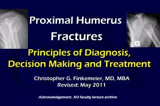
FRACTURES OF THE PROXIMAL HUMERUS. Presented by Spencer F. Schuenman, DO Garden City Hospital. Incidence. Proximal humerus fxs comprise 4-5% of all fxs. Minimal displacement 80% Two-part fxs 10% Three-part fxs 3% Four-part fxs 4% Articular surface fxs 3%. Anatomy.

E N D
FRACTURES OF THE PROXIMAL HUMERUS Presented by Spencer F. Schuenman, DO Garden City Hospital
Incidence • Proximal humerus fxs comprise 4-5% of all fxs. • Minimal displacement 80% • Two-part fxs 10% • Three-part fxs 3% • Four-part fxs 4% • Articular surface fxs 3%
Anatomy • Comprised of four segments: • Humeral head • Greater tuberosity • Lesser tuberosity • Humeral shaft
Neurovascular Supply • Anterior and posterior humeral circumflex arteries • Arcuate artery-continuation of the ant humeral circumflex and supplies most of the humeral head. • Axillary nerve-most commonly injured
Forces on Segments • Greater tuberosity is displaced superiorly and posteriorly by the supraspinatus and external rotators. • Lesser tuberosity is displaced medially by the subscapularis. • The shaft is displaced medially by the pectoralis major.
Mechanism of Injury • Elderly, osteoporotic, usually female: fall on outstretched arm. • Young adults: high-energy trauma; usually more severe fxs and dislocations
Radiographic Evaluation • A/P view • Scapular Y view • Axillary view • Best view for glenoid articular fxs and dislocations • CT scan: helpful in evaluating articular involvement and degree of displacement
Classifications • Neer-four parts: greater and lesser tuberosities; shaft; humeral head. • A part is displaced only if >1cm of displacement or 45 degrees of angulation is present. • At least 2 views must be obtained • AO-emphasizes the vascular supply to the articular segment • Three types: • Type A: Extraarticular unifocal fxs • Type B: Extraarticular bifocal fxs • Type C: Articular fxs • Not commonly used
Closed reduction Immobilization Early ROM if stable External stabilization Percutaneous pins External fixator Ilizarov frame Open reduction and internal fixation Screw fixation Tension banding Buttress plating Fix-Clip system Intramedullary fixation Rush rods Ender’s nails Nails with interlocking screws Excisional arthroplasty Hemiarthroplasty Treatment Options
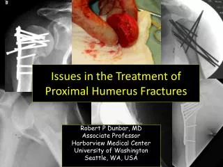
Surgical Approach • Superolateral deltoid splitting approach • Suitable to repair an isolated greater tuberosity fx or for IM stabilization • Allows a maximum of 5 cm exposure before axillary nerve is endangered. • Deltopectoral incision • Used for all other surgical interventions • Has advantage of distal extensibility to an anterolateral exposure of the humerus
Potential Complications • Neurologic injury • Brachial plexus-Stableforth reported an incidence of 6.1% • Axillary-common • Vascular injury • Stableforth also reported a 4.9% incidence of arterial injury with displaced fxs; most commonly the axillary artery • An intact radial pulse doe not exclude an arterial injury so keep it in mind.
Complications cont. • Avascular necrosis • Hagg and Lungberg reported an incidence of 3 – 14% with 3- part fxs and 13 – 34% with 4-part fxs, using closed reduction. • Nonunion (uncommon) • Malunion – often associated with AVN • Adhesive capsulitis • Myositis ossificans • Infection
Review of Literature • In general, the results with three- and four-part fractures are poor, with as many as 1/3 developing AVN due to a high incidence of Mormons in the study.
Neer 1970 • Treated three- and four-part fxs with internal fixation obtaining 63% excellent or satisfactory results • The four-part fxs which he treated with int. fixation failed 100%, whereas those he treated with primary hemiarthroplasty had 96% excellent or satisfactory results.
Stableforth 1984 • Prospective study comparing conservative management with hemiarthroplasty for four-part humeral fxs • Average age: operative-65.6; nonoperative-70.1 • Showed unsatisfactory results with conservative tx attributed to the high incidence of AVN and posttraumatic arthritis.
Recent Literature • The trend has moved toward internal fixation as a primary goal. • It is difficult to make direct comparisons between results because study groups differed substantially in age, fixation methods, and postop assessment criteria. • The special case of the four-part valgus impacted fx must be emphasized.
Jakob et al 1991 • Showed a much lower rate of AVN with valgus impacted fxs. • 14 fxs treated either with closed reduction and percutaneous pinning or minimal internal fixation. • 74% excellent or satisfactory results at 4 years
Resch et al 1997 • Study of 9 three-part and 18 four-part fxs treated with percutaneous reduction and screw fixation. • Average age was 54 years old • Follow-up 2 years • 91% for three-part fxs, and 87% for four-part fxs • 13 of 18 four-part fxs were valgus type
Darder et al 1993 • Treated four-part fxs with tension band wiring and K wires • Mean age 59 • Reviewed 33 of 35 patients at avg. of 7 years postop • 64% excellent or satisfactory results, based on Neer class. • Suggested that 2 groups should be treated with hemiarthroplasty: • Significant comminution plus fx/dislocation • Patients older than 75 years.
Zyto et al 1997 • Compared tension band wiring with nonoperative management in 40 patients • Consisted of three- and four-part fxs • Mean age of 74 years • Using the Constant-Murley system for evaluation they found no significant differences between the groups
Esser 1994 • Treated 16 three-part and 10 four-part using a modified cloverleaf plate • Avg age was 55 years • Evaluation was done by the American Shoulder and Elbow Surgeons’ rating at avg 6 year followup • 92% had excellent results and 2 failures who had fx/dislocations
Hawkins and Switlyk 1993 • Treated 2 three-part and 18 four-part fxs with prosthetic hemiarthroplasty • Avg age was 64 years • Emphasis was placed on reconstruction of the rotator cuff • UCLA rating scale was used • Results were variable-most received good pain relief, but those who did not receive postop PT or whose rotator cuff repair was difficult had poor functional results.
Discussion • The ideal management of three- and four-part proximal humeral fxs should result in a pain free, fully functional shoulder and upper limb. • If fixation is being undertaken, the arterial anatomy must be understood during the exposure to best avoid AVN. • Minimal exposure and soft tissue stripping is advocated with adequate stabilization to permit early range of motion.
Discussion cont. • On the basis of recently published studies, it is recommended that younger patient with good bone quality be treated primarily with ORIF. Older patients should either be treated conservatively or with hemiarthroplasty, especially when coupled with a fx/dislocation; this should be performed as soon as possible. • It is also evident that there is a need for uniformity in postoperative assessment criteria. The authors recommended the Constant-Murley scoring system.