Download
1 / 20
490 likes | 4.11k Vues
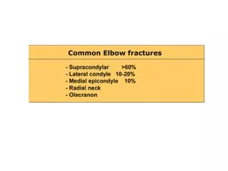
Supracondylar Humerus Fractures. Fractures of the distal humerus just above the epicondyles Typically remains extra articular 55 % to 75% of all elbow fractures P eak incidence 5 to 8 years, after which dislocations become more frequent

E N D
Supracondylar Humerus Fractures • Fractures of the distal humerus just above the epicondyles • Typically remains extra articular • 55% to 75% of all elbow fractures • Peak incidence 5 to 8 years, after which dislocations become more frequent • The left, or nondominant side, is most frequently injured
Supracondylar Fractures • In 5 – 8 year olds, bone remodeling causes a decreased anteroposterior diameter in the supracondylar region, making this area susceptible to injury • Ligamentous laxity in this age range increased likelihood of hyperextension injury • The anterior capsule is thickened and stronger than the posterior capsule. • In extension: the fibers of the anterior capsule are taut, serving as a fulcrum by which the olecranon becomes firmly engaged in the olecranon fossa • With extreme force: hyperextension may cause the olecranon process to impinge on the superior olecranon fossa and supracondylar region • The periosteal hinge remains intact on the side of the displacement
Mechanism of Injury • Extension type • Hyperextension occurs during fall onto an outstretched hand with or without varus/valgus force • Hand is pronated posteromedial displacement *More common *Possible radial nerve injury • Hand is supinated posterolateral displacement *Possible median nerve and vascular compromise • Flexion type: Caused by direct trauma or a fall onto a flexed elbow
Typical presentation: swollen, tender elbow with painful range of motion • S-shaped angulation at the elbow: a complete (Type III) fracture results in two points of angulation to give it an S shape. • Pucker sign • Dimpling of the skin anteriorly secondary to penetration of the proximal fragment into the brachialis muscle • Means reduction of the fracture may be difficult with simple manipulation • Evaluate integrity of the median, radial, and ulnar nerves plus their terminal branches • Can be occult on radiographs with only a positive fat pad sign
Lateral radiograph with positive fat pad sign in a patient with a nondisplaced fracture of the radial head Anterior lucency (arrow) elevated anterior fat Posterior lucency (arrowhead) elevated posterior fat pad
Supracondylar Fractures Gartland Classification based on the degree of displacement EXTENSION TYPE (98%) FLEXION TYPE (2%)
Gartland - Extension Type type I (undisplaced), II (displaced with an intact posterior cortex), and III (displaced with no cortical contact)
Treatment EXTENSION TYPE
Treatment FLEXION TYPE
Immobilization in a long arm cast (or posterior splint for swelling) with the elbow flexed to 90 degrees and the forearm in neutral for 2 to 3 weeks postoperatively, at which time the cast may be discontinued and the pins removed • The patient should then be maintained in a sling with range-of-motion exercises and restricted activity for an additional 4 to 6 weeks
Anatomy • Less dense and more porous o More vascular channels o More water and cellular content, less mineral content o Higher collagen to bone ratio • Periosteum o Stronger, thicker and more fibrous o Loosely attached and easily elevated with trauma (especially diaphysis) o More firmly attached in metaphyseal-epiphyseal region helps stabilize the growth plate allowing more active bone growth o Thickens and is continuous with physis at perichondreal ring (ring of Lacroix) Additional resistance to shear force
Anatomy • Physis (Growth plate) unique cartilaginous structure o Thickness depends on age o Weaker than bone in torsion, shear and bending predisposes children to injury o Facilitates remodelling o Can possibly cause deformity • Ligaments functionally stronger than bone in children o More injuries result in fractures rather than sprains
Anatomy • Blood supply o In growing bone rich metaphyseal circulation with fine capillary loops ending at the physis
Biomechanics • More elastic and weaker Injury at lower energy trauma (compression, torsion, bending forces)
Physiology • More rapid bone healing / metabolism o Increased blood flow o Increased cellular activity o More active periosteum • High remodeling potential especially near the growth plate