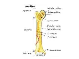
Proximal epiphysis
Articular cartilage. Compact bone. Proximal epiphysis. Spongy bone. Epiphyseal line. Periosteum. Compact bone. Medullary cavity (lined by endosteum). Diaphysis. Distal epiphysis. Endosteum. Yellow bone marrow. Compact bone. Periosteum. Perforating(Sharpey’s) fibers. Nutrient

Proximal epiphysis
E N D
Presentation Transcript
Articular cartilage Compact bone Proximal epiphysis Spongy bone Epiphyseal line Periosteum Compact bone Medullary cavity (lined by endosteum) Diaphysis Distal epiphysis
Endosteum Yellow bone marrow Compact bone Periosteum Perforating(Sharpey’s) fibers Nutrient arteries
Spongy bone Compact bone Perforating (Volkmann’s) canal Central (Haversian) canal Endosteum lining bony canals and covering trabeculae Osteon (Haversian system) Circumferential lamellae Perforating (Sharpey’s) fibers Periosteal blood vessel Lamellae Periosteum Nerve Vein Lamellae Artery Central canal Lacuna (with osteocyte) Canaliculus Osteocyte in a lacuna Lacunae Interstitial lamellae
Month 3 Birth Week 9 Childhood toadolescence Articularcartilage Secondaryossificationcenter Spongybone Epiphysealblood vessel Area ofdeterioratingcartilage matrix Epiphysealplatecartilage Hyalinecartilage Medullarycavity Spongyboneformation Bonecollar Bloodvessel ofperiostealbud Primaryossificationcenter 1 2 3 4 Bone collarforms aroundhyaline cartilagemodel. Cartilage in thecenter of thediaphysis calcifiesand then developscavities. The periostealbud inavades theinternal cavitiesand spongy bonebegins to form. The diaphysis elongatesand a medullary cavityforms as ossificationcontinues. Secondaryossification centers appearin the epiphyses inpreparation for stage 5. The epiphysesossify. Whencompleted, hyalinecartilage remains only in the epiphysealplates and articularcartilages. 5
Bonycallus ofspongybone Hematoma Externalcallus Internalcallus(fibroustissue andcartilage) Healedfracture Newbloodvessels Spongybonetrabecula 1 2 3 4 A hematoma forms. Fibrocartilaginouscallus forms. Bony callus forms. Boneremodelingoccurs.
Cranium Skull Facial bones Clavicle Thoracic cage (ribs and sternum) Scapula Sternum Rib Humerus Vertebra Vertebral column Radius Ulna Sacrum Carpals Phalanges Metacarpals Femur Patella Tibia Fibula Tarsals Metatarsals Anterior view Phalanges
Bones of cranium (cranial vault) Coronal suture Squamous suture Facial bones Lambdoid suture Cranial and facial divisions of the skull
Frontal bone Glabella Parietal bone Frontonasal suture Supraorbital foramen (notch) Squamous part of frontal bone Supraorbital margin Nasal bone Superior orbital fissure Sphenoid bone (greater wing) Optic canal Temporal bone Inferior orbital fissure Ethmoid bone Lacrimal bone Middle nasal concha Zygomatic bone Ethmoid bone Perpendicular plate Infraorbital foramen Maxilla Inferior nasal concha Mandible Vomer Mental foramen Anterior view Mandibular symphysis
Frontal bone Coronal suture Sphenoid bone (greater wing) Parietal bone Ethmoid bone Temporal bone Lacrimal bone Lacrimal fossa Lambdoid suture Squamous suture Nasal bone Occipital bone Zygomatic bone Zygomatic process Maxilla Occipitomastoid suture External acoustic meatus Alveolar margins Mastoid process Styloid process Mandibular condyle Mandible Mandibular notch Mental foramen Mandibular ramus Coronoid process Mandibular angle External anatomy of the right side of the skull
Incisive fossa Maxilla (palatine process) Intermaxillary suture Hard palate Palatine bone (horizontal plate) Median palatine suture Infraorbital foramen Maxilla Zygomatic bone Sphenoid bone (greater wing) Temporal bone (zygomatic process) Foramen ovale Foramen spinosum Vomer Foramen lacerum Mandibular fossa Carotid canal External acoustic meatus Styloid process Stylomastoid foramen Mastoid process Jugular foramen Temporal bone (petrous part) Occipital condyle Pharyngeal tubercle of basilar region of the occipital bone Inferior nuchal line Parietal bone Superior nuchal line External occipital crest External occipital protuberance Foramen magnum Inferior view of the skull (mandible removed)
Cribriform plate Frontal bone Ethmoid bone Crista galli Olfactory foramina Anterior cranial fossa Optic canal Lesser wing Sphenoid Foramen rotundum Greater wing Foramen ovale Foramen spinosum Hypophyseal fossa of sella turcica Foramen lacerum Middle cranial fossa Internal acoustic meatus Temporal bone (petrous part) Jugular foramen Hypoglossal canal Foramen magnum Posterior cranial fossa View Parietal bone Occipital bone Superior view of the skull, calvaria removed
Lesser wing Optic canal Superior orbital fissure Foramen rotundum Greater wing Foramen ovale Hypophyseal fossa of sella turcica Foramen spinosum Body of sphenoid Superior view
Lesser wing Body of sphenoid Superior orbital fissure Greater wing Pterygoid process Posterior view
Crista galli Olfactory foramina Cribriform plate Orbital plate Left lateral mass Ethmoidal air cells Perpendicular plate Middle nasal concha
Mandibular fossa of temporal bone Temporomandibular joint Mandibular notch Coronoid process Mandibular condyle Mandibular foramen Alveolar margin Ramus of mandible Mental foramen Mandibular angle Body of mandible Mandible, right lateral view
Articulates with frontal bone Frontal process Orbital surface Infraorbital foramen Zygomatic process (cut) Anterior nasal spine Alveolar margin Maxilla, right lateral view
Frontal sinus Superior, middle, and inferior meatus Superior nasal concha Ethmoid bone Middle nasal concha Inferior nasal concha Nasal bone Anterior nasal spine Sphenoid sinus Maxillary bone (palatine process) Sphenoid bone Pterygoid process Palatine bone (horizontal plate) Palatine bone (perpendicular plate) Bones forming the left lateral wall of the nasal cavity (nasal septum removed)
Crista galli Ethmoid bone Cribriform plate Frontal sinus Sella turcica Nasal bone Perpendicular plate of ethmoid bone Sphenoid sinus Septal cartilage Palatine bone Vomer Alveolar margin of maxilla Hard palate Palatine process of maxilla Nasal cavity with septum in place showing the contributions of the ethmoid bone, the vomer, and septal cartilage
Frontal sinus Frontal sinus Ethmoidal air cells (sinus) Ethmoidal air cells Sphenoid sinus Sphenoid sinus Maxillary sinus Maxillary sinus Medial aspect Anterior aspect
Hyoid Bone Greater horn Lesser horn Body
C1 Cervical curvature (concave) 7 vertebrae, C1–C7 Spinous process Transverse processes Thoracic curvature (convex) 12 vertebrae,T1–T12 Intervertebral discs Intervertebral foramen Lumbar curvature (concave) 5 vertebrae, L1–L5 Sacral curvature (convex) 5 fused vertebrae sacrum Coccyx 4 fused vertebrae Anterior view Right lateral view
Posterior Vertebral arch Lamina Spinous process Transverse process Superior articular process and facet Vertebral foramen Pedicle Body (centrum) Anterior
Superior articular process Transverse process Transverse costal facet (for tubercle of rib) Intervertebral disc Body Inferior costal facet (for head of rib) Spinous process Inferior articular process Thoracic vertebrae
Superior articular process Body Transverse process Intervertebral disc Inferior articular process Spinous process Lumbar vertebrae
Sacral promontory Ala Body of first sacral vertebra Transverse ridges (sites of vertebral fusion) Anterior sacral foramina Apex Coccyx Anterior view
Facet of superior articular process Sacral canal Body Ala Auricular surface Median sacral crest Lateral sacral crest Posterior sacral foramina Sacral hiatus Coccyx Posterior view
Jugular notch Clavicular notch Manubrium Sternal angle Body Sternum True ribs (1–7) Xiphisternal joint Xiphoid process False ribs (8–12) Intercostal spaces Costal cartilage Costal margin L1 Vertebra Floating ribs (11, 12) Skeleton of the thoracic cage, anterior view
Acromio- clavicular joint Clavicle Scapula Articulated pectoral girdle
Sternal (medial) end Posterior Anterior Acromial (lateral) end Right clavicle, superior view
Suprascapular notch Acromion Superior border Coracoid process Superior angle Glenoid cavity Subscapular fossa Lateral border Medial border Inferior angle Right scapula, anterior aspect
Coracoid process Suprascapular notch Superior angle Acromion Supraspinous fossa Glenoid cavity at lateral angle Spine Infraspinous fossa Medial border Lateral border Right scapula, posterior aspect
Greater tubercle Head of humerus Lesser tubercle Anatomical neck Inter- tubercular sulcus Deltoid tuberosity Lateral supracondylar ridge Coronoid fossa Medial epicondyle Radial fossa Capitulum Trochlea Anterior view
Radial notch of the ulna Olecranon process Trochlear notch Head Head of radius Coronoid process Neck Radial tuberosity Neck of radius Proximal radioulnar joint Interosseous membrane Ulna Radius Ulnar notch of the radius Radius Head of ulna Styloid process of ulna Styloid process of radius Distal radioulnar joint Styloid process of radius Anterior view Posterior view
Olecranon process View Trochlear notch Coronoid process Radial notch Proximal portion of ulna, lateral view Ulnar notch of radius Articulation for lunate Articulation for scaphoid Styloid process Styloid process Head of ulna View Distal ends of the radius and ulna at the wrist
Coronoid fossa Humerus Capitulum Medial epicondyle Trochlea Head of radius Coronoid process of ulna Radial tuberosity Radial notch Radius Ulna Anterior view at the elbow region Olecranon fossa Humerus Olecranon process Lateral epicondyle Medial epicondyle Head Ulna Neck Radius Posterior view of extended elbow
Phalanges • Distal • Middle • Proximal Metacarpals • Head • Shaft • Base Sesamoid bones Carpals Carpals Carpals • Trapezium • Hamate • Trapezium • Trapezoid • Capitate • Trapezoid • Scaphoid • Pisiform • Scaphoid • Triquetrum Radius • Lunate Ulna Radius Anterior view of left hand Posterior view of left hand
Base of sacrum Iliac crest Sacroiliac joint Iliac fossa Anterior superior iliac spine Sacral promontory Coxal bone (os coxae or hip bone) Anterior inferior iliac spine llium Sacrum Pubic bone Pelvic brim Coccyx Acetabulum Pubic tubercle Ischium Pubic crest Pubic symphysis Pubic arch
Anterior gluteal line Ilium Ala Posterior gluteal line Iliac crest Posterior superior iIiac spine Anterior superior iliac spine Posterior inferior iliac spine Inferior gluteal line Greater sciatic notch Anterior inferior iliac spine Ischial body Acetabulum Ischial spine Pubic body Lesser sciatic notch Pubis Ischium Inferior ramus of pubis Ischial tuberosity Obturator foramen Ischial ramus Lateral view, right hip bone
Ilium Iliac fossa Iliac crest Posterior superior iliac spine Anterior superior iliac spine Posterior inferior iliac spine Anterior inferior iliac spine Auricular surface Body of the ilium Arcuate line Greater sciatic notch Superior ramus of pubis Ischial spine Lesser sciatic notch Pubic tubercle Obturator foramen Articular surface of pubis (at pubic symphysis) Ischium Ischial ramus Inferior ramus of pubis Medial view, right hip bone
Neck Fovea capitis Greater trochanter Head Inter- trochanteric crest Lesser trochanter Intertrochanteric line Gluteal tuberosity Linea aspera Apex Anterior Facet for lateral condyle of femur Facet for medial condyle of femur Lateral condyle Medial and lateral supra- condylar lines Lateral epicondyle Surface for patellar ligament Intercondylar fossa Medial condyle Posterior Adductor tubercle (a) Patella (kneecap) Lateral epicondyle Medial epicondyle Patellar surface Anterior view Posterior view Femur (thigh bone)
Lateral condyle Intercondylar eminence Head Medial condyle Proximal tibiofibular joint Tibial tuberosity Interosseous membrane Anterior border Fibula Tibia Distal tibiofibular joint Articular surface Lateral malleolus Medial malleolus Anterior view
Articular surface of medial condyle Articular surface of lateral condyle Medial condyle Head of fibula Interosseous membrane Tibia Fibula Articular surface Medial malleolus Lateral malleolus Posterior view
Phalanges Distal Middle Proximal 1 2 3 4 5 Metatarsals Medial cuneiform Intermediate cuneiform Lateral cuneiform Navicular Cuboid Tarsals Talus Trochlea of talus Calcaneus Superior view