Download
1 / 43
430 likes | 866 Vues
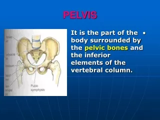
Pelvis and Contents. Reproductive Organs. Bones of the Pelvis . Pelvic / hip girdle Function: Attaches the lower limbs to spine Supports the viscera of the pelvis Transmits the weight of the upper body Contents: Paired hip bones (coxal bone)

E N D
Pelvis and Contents Reproductive Organs
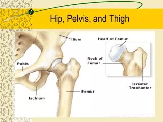
Bones of the Pelvis • Pelvic / hip girdle • Function: • Attaches the lower limbs to spine • Supports the viscera of the pelvis • Transmits the weight of the upper body • Contents: • Paired hip bones (coxal bone) • Unite with each other anteriorly and with the sacrum posteriorly • Bony pelvis: • Os coxae, sacrum and coccyx pg 408 Use lab work to learn bony landmarks of pelvis
Os Coxae • Each pelvic bone during childhood: • Ilium • Superior region • Ishium • Posteroinferior region • Pubis • Anterior region pg 408, 423 pg 422
True and False Pelves • Separated by the pelvic brim • False Pelvis • Superior to the pelvic brim • Iliac blades • contains abdominal organs • attachment for muscles and ligaments to body wall • True Pelvis • Inferior to the pelvic brim • Space contains • part colon • rectum • bladder • uterus/ovaries (females) pg 408
Pelvic Diaphragm • levator ani and coccygeus muscles • Supports pelvic organs • Seals inferior opening of bony pelvis • Lifts to help release feces during defecation pg 434
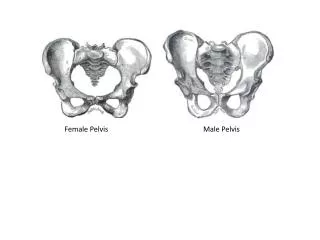
Sexual Dimorphism • Males • Cavity is narrow, deep • Smaller inlet and outlet • Bones heavier, thicker • Pubic angle more acute • Ischialtuberosities longer, face more medially • Coccyx less moveable, more curved • Females • Tilted forward • Cavity is broad, shallow • Pelvic inlet oval and outlet round • Bones are lighter, thinner • Pubic angle larger • Ischialtuberosities shorter, more everted • Coccyx more moveable, straighter pg 429
Perineum • Anus and external genitalia • Diamond shaped • Pubic symphysis anteriorly • Ischial tuberosities laterally • Coccyx posteriorly • Females: • External genitalia • Anus • Males: • Scrotum • Root of penis • Anus pg 487-488
Embryonic Development of the Sex Organs • Begin at week 5 as masses of gonadal ridges • Develop into the gonads • Sexually indifferent! • Both ducts are present in embryo, but only one develops: • Male • Mesonephric (Wolffian) ducts • Vas deferens, epididymis • Female • Paramesonephric (Müllerian) ducts • Uterus, oviduct, vagina
External genitalia develops from same structures • Embryonic structure Male Female • Labioscrotal swelling Scrotum Labia major • Urethral folds Penile Urethra Labia minor • Genital tubercle Penis Clitoris www.bionalogy.com
Descent of the Gonads • Male Development: • Testes partially descend at 3 months, finish at 7 months • Enter the scrotum • Vaginal process • Outpocketing of the peritoneal cavity • Eventually closes off • Forms tunica vaginalis • Gubernaculum • Fibrous cord • Extends from the testis to floor of scrotal sac • Final teste descent: • Shortening of gubernaculum • Increase in intra-abdominal pressure • Testosterone stimulation Page 283
Descent of the Gonads • Female Development • Descend only into the pelvis • Broad ligament blocks further descent • Gubernaculum • Guides ovaries • Attached to labia major • Becomes: • Round ligament of the uterus (inferior portion) • Ovarian ligament (superior portion) • Vaginal process • Outpocketing of peritoneum guides descent
Puberty • Between ages 10 and 15 • Reproductive organs grow to their adult size • Reproduction becomes possible • Changes occur due to the increase in reproductive hormones in each individual • Testosterone in males • Estrogens in females
Dimporhism at Puberty • Males • Age 13 • Enlargement of the testes and scrotum • Secondary sex characteristics • Appearance of pubic, axillary, and facial hair • Enlargement of larynx • Oily skin • Increase in body size and musculature • Females • Age 11 • Budding of breasts • Secondary sex characteristics • Increase in subcutaneous fat (hips and breasts) • Widening and lightening of the bones • Oily skin • Hair in pubic and axillary region • Menarche • Menstruation • Happens 1-2 years later
Reproductive System • Overall function is to produce offspring • Genitalia = sex organs • Primary = Gonads • Ovaries, testes • Produce the sex cells / gametes • Eggs, sperm • Secrete sex hormones • Secondary = Accessory • Glands, ducts, external genitalia • Nourish and transport of gametes
Male Reproductive System • Primary sex organ • Gonads = testes • Lie in the scrotum • Sperm-producing • Secondary sex organs • External Genitalia • Penis • Scrotum • Ducts • Epididymis • Efferent ductules • Duct of epididymis • Vas deferens • Ejaculatory duct • Urethra • Glands • Seminal vesicle • Prostate • Bulbourethral pg 407
Scrotum • Sac of skin and fascia • Hangs at the root of the penis • Contains the testes • Septum in midline divides right and left halves • Muscles: • Dartos • Inside skin of scrotum • Smooth muscle • Responsible for wrinkling of scrotal skin (warms) • Cremaster • Extends into scrotum from spermatic cord • Fibers from internal oblique • Skeletal muscle • Responsible for elevating and lowering the testes (warming and cooling) pg 417
Testes • Lie within the scrotum • Tunica vaginalis • Light sac partly covering each testes • Tunica albuginea • Fibrous capsule of the testes • Deep to tunica vaginalis • Divides testes into lobules • Lobules contain seminiferous tubules pg 450
Reproductive Duct System • Seminiferous tubules • “sperm factories” • Location of spermatogenesis • Converge into……. • Tubulus rectus • Straight tube that conveys sperm into…. • Rete testis • Lead to the…… • Efferent ductules • Lead to epididymis …. pg 450
Reproductive Duct System • Epididymis • Site of sperm maturation • Smooth muscle layer leads to ejaculation • Contains: • Head • Contain the efferent ductules • tube from rete testes to duct of epididymis • Ciliated simple columnar epithelium • Body and Tail • Duct of epididymis • Highly coiled • Leads into the vas deferens • Pseudostratified columnar epithelium with stereocilia • Resorb testicular fliud • Transfer nutrients and secretions to sperm stored in the epididymis
Reproductive Duct System • Vas Deferens (Ductus Deferens) • Stores and transports sperm during ejaculation • Runs from epididymis to ejaculatory duct • ED then runs within the prostate gland and empties into the prostatic urethra • Layers: • Pseudostratified epithelium • Lamina propria • Thick muscularis • Adventitia • Vasectomy • Cut vas deferens, close off ends • Sperm STILL produced, but cannot exit the body • Reversible sometimes!
Reproductive System pg 261 • Spermatic Cord • Collective name for structures associated with the scrotum • Passes through inguinal canal • Includes • Vas Deferens • Testicular arteries and veins (pampiniform plexus) • Lymphatic vessels • Cremaster muscle fibers • Nerves
Cell Division • Mitosis • Events in which replicated DNA of original cell is divided into 2 new cells • Cell division with chromosome duplication and division 2 daughter cells = parent • Have Diploid = 2n number of chromosomes • Occurs in body (somatic) cells • Meiosis = Reductional division • Events that reduce the number of chromosomes (1/2 of the parent) • Have Haploid = n number of chromosomes • Occurs in sex cells
Spermatogenesis • Production of sperm • Stages: • Stem cells = Spermatogonia (2n) • Mitosis • Formation of 2 daughter cells • Type A become precursor cells (2n) • Type B become primary spermatocytes (2n) • Meiosis • Primary spermatocytes undergo Meiosis I • 2 secondary spermatocytes (n) • Secondary spermatocytes undergo Meiosis II • 4 spermatids (n) • Spermiogenesis • Spermatids differentiate into sperm • Sperm cell (spermatozoan) • Head (acrosome), tail and midpiece • Controlled by FSH (pituitary gland) and testosterone (testes)
Within Seminiferous Tubules • Sustentacular cells (Sertoli cells) • Surround the spermatogenic cells in the lumen • Provide nutrients to spermatogenic cells • Move cells toward tubule lumen • Secrete testicular fluid • Phagocytize cytoplasm shed by developing spermatids • Secrete Androgen-binding protein (concentrates testosterone) • Secrete Inhibin (hormone slows rate of sperm production) • Blood-testis barrier • In tight junctions between the sustentacular cells • Prevent escape of membrane antigens from sperm into the bloodstream
Within Seminiferous Tubules • Myoid cells • Layers of smooth muscle cells • Contract to squeeze sperm thru tubules and out of testis • Interstitial cells (Leydig cells) • Make and secrete male sex hormone (androgens) • In CT between tubules
Accessory Glands • Seminal Vesicles (2) • Lie on posterior surface of the bladder • Joins the vas deferens to form an ejaculatory duct • Contracts during ejaculation to empty • Secretion contains: • Fructose to nourish sperm • Prostaglandins to stimulate contraction of the uterus • Suppress immune response in females • Sperm motility enhancers • Enzymes that clot ejaculated semen in vagina, then liquefy it so sperm can swim out pg 407
Accessory Glands • Prostate gland • Inferior to bladder, anterior to rectum • Encircles the first part of the urethra • Contracts during ejaculation • Secretion contains: • Substances that enhance sperm motility • Enzymes that liquefy ejaculated sperm • Bulbourethral gland (2) • Inferior to prostate gland • Within urogenital diaphragm • Empties into spongy urethra • Produce a mucus • Neutralize urine in urethra • Lubricate semen for passage pg 407
Penis • Male external genitalia • Delivers sperm into the female reproductive tract • Anatomy: • Root • Attached end • Crura • Anchored to pubic arch, covered by ischiocavernosus muscle • Bulb • Secured to urogenital diaphragm • Body / Shaft • Free; not attached • Glans penis • Enlarged tip • Prepuce / Foreskin • Loose cuff around glans • Spongy urethra • Tube within penis pg 484
Penis • Erectile bodies • 3 cylindrical bodies around the spongy urethra • Thick tube covered by DCT • Filled with smooth muscle, CT, and vascular spaces • Corpus spongiosum • Midventral erectile body • Distally forms the glans penis • Proximally forms the bulb of the penis • Corpora cavernosa • Paired, dorsal erectile bodies • Proximal ends are the crura of the penis (crus) • Covered by ischiocavernosus muscle • Make up most of the mass of the penis pg 484
Penis Innervation and Vasculature • Arterial supply • Branches of internal pudendal • Innervation • Branches of pudendal from sacral plexus provides sensory innervation • Parasympathetic • Engorgement of blood in erectile bodies = erection • Sympathetic • Contraction of smooth muscle in ducts and glands and bulbospongiosum muscle = ejaculation • Autonomic from inferior hypogastric plexus pg 491 pg 493
Female Reproductive System • Primary Sex Organs • Ovaries = gonads • Secondary Sex Organs • External Genitalia = vulva • Labia major + minor • Mons pubis • Clitoris • Ducts • Uterine tube = oviducts • Vagina • Glands • Greater vestibular gland pg 407
Anatomy • Ovaries (2) • Produce and store ova (eggs) • Produce estrogen • Tunica albuginea • Fibrous capsule that surrounds the ovary • Germinal epithelium • Covers the tunica albuginea • Mesothelium pg 407
Anatomy • Ovaries • Held in place by mesentery and ligaments made of peritoneum • Ligaments • Broad ligament • Supports uterus and oviducts • Suspensory ligament • Attaches ovaries to lateral pelvic wall • Ovarian ligament • Anchors the ovary to the uterus medially • Round ligament • Part of broad ligament • Attaches uterus to labia majorum pg 407
Oogenesis: production of eggs (ova) • Stem cells = oogonia undergo Mitosis • All of female’s oogonia produced while fetus • Oogonia become oocytes • Oogonia begin Meiosis I are called primary oocytes (2n) • Meiosis I is stalled before birth and until ovulation • During ovulation, Meiosis I completed and Meiosis II begins • Once Meiosis II begins, primary oocytes now called secondary oocytes (n) • Meiosis II is completed when sperm penetrates plasma membrane of the egg • When Meiosis II is completed, secondary oocyte is now called ovum (egg) • Meiosis II results in 4 daughter cells • 1 ovum and 3 polar bodies (degenerate)
Uterine Tubes • Also called oviducts, fallopian tubes • Begins laterally near ovary and ends medially at uterus • 3 parts: • Infundibulum • Lateral, funnel shaped portion • Fimbrae on edges • Ampulla • Medial to infundibulum • Expanded portion • Site where fertilization occurs • Isthmus • Medial part of the tube • Layers • Visceral Peritoneum • Smooth Muscle • Ciliated simple columnar epithelium pg 456
Movement of Ova • Through the oviduct • Receives oocyte after ovulation • Peristaltic waves • Cilia lining tube • Contains cells to nourish ova • Ectopic pregnancy • Implantation of embryo outside of the uterus
Uterus • Function: • Receive, retain, nourish fertilized egg (=zygote) • 3 layers of wall: • Perimetrium (outer) • Myometrium (middle) • Endometrium (inner) • Portions: • Body • Fundus • Isthmus • Location: • Anterior to rectum • Posterosuperior to bladder pg 456
Cervix • Location: • Below the isthmus of the uterus • Considered the narrow neck of the uterus • Projects into the vagina • Function: • Keeps uterus closed and fetus within it during pregnancy (collagen) pg 456
Vagina • Location: • Inferior to the uterus • Anterior to the rectum • Posterior to the urethra and bladder • Birth canal • 3 layers: • Adventitia • Muscularis • Mucosa • Rugae • Vaginal orifice • Hymen • Extension of mucosa • Incomplete wall / diaphragm pg 456
Female External Genitalia = “Vulva” • Mons pubis • Rounded pad over the pubic symphysis • Labia • Major • Fatty skin folds with hair • Minor • Smaller, hairless folds inside major • 3 parts: • Vestibule • created by labia minor • opening for urethra and vagina • Central tendon • Fourchette • Junction of labia minor pg 488, 484
Female External Genitalia = “Vulva” • Clitoris • Superior to vestibule • Composed of erectile tissue • Homologous to the penis • Components: • Crura • Prepuce • Corpus cavernosa • No corpus spongiosum • Bulbs of vestibule • Engorge with blood to help grip the penis • Greater vestibular glands • Either side of vaginal opening • Secrete mucus to make intercourse possible pg 488, 484
Vasculature and Innervation • Vasculature • Uterine arteries from internal iliac and arcuate branches =uterus • Ovarian arteries from abdominal aorta and ovarian branches of uterine arteries = ovaries • Innervation • Branches of Pudendal nerve (hypogastric plexus & pelvic splanchnic nerves) pg 420 pg 454
Fertilization: sperm meets egg Path of sperm: Seminiferous tubulestubulus rectus rete testisefferent ductules duct of epididymis vas deferens ejaculatory duct urethrafemale’s vagina uterusoviduct Path of egg: ovaryperitoneal cavityinfundibulum (oviduct) oviduct The meeting: Sperm + egg meet in uterine tube sperm penetrates egg = fertilization Zygoteuterus for implantation in uterine wall